

 PDF
PDF ePub
ePub Citation
 Print
Print


Abstract
Objective
This study was to change of pulp blood flow among maxillary and mandibular anterior tooth with mild crowding and adjacent teeth using Ultrasound Doppler graphy.
Methods
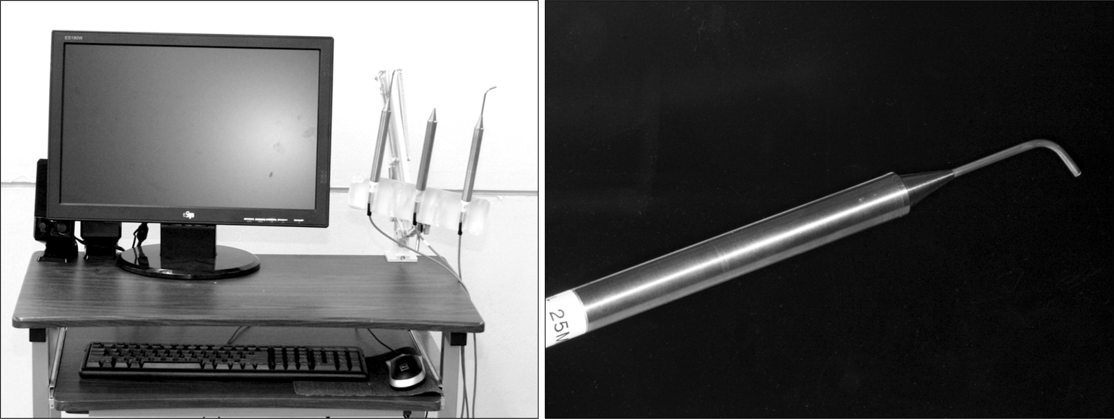
The change of pulp blood flow was measured three times using Ultrasound Doppler graphy; before the attachment of brackets, after 3 week, and after 6 week. The sample consists of 15 year old eighteen patients.
Results
Before the attachment of brackets, after 3 weeks, and after 6 weeks, there were no significant differences in the change of pulp blood flow in each part (maxilla and mandible) and each tooth according to period. In addition, to compare internal dangerousness of loss of the pulp vitality, when pulp blood flow is compared in each tooth before orthodontic treatment, there were no statistically significant differences in maxillary lateral incisor and mandibular canine but it showed low values in all measurement items (p > 0.05).
Go to : 
REFERENCES
1.Min JH., Cho JH., Lee KH., Hwang HS. The effects of ipriflavone on the periodontal reorganization following experimental tooth movement in the rat. Korean J Orthod. 2008. 38:347–57.

2.Proffit WR. Contemporary orthodontic. 4th ed.St Louis: Mosby;2008. p. 335–51.
3.Anstendig HS., Kronman JH. A histologic study of pulpal reaction to orthodontic tooth movement in dogs. Angle Orthod. 1972. 42:50–5.
4.Hamersky PA., Weimer AD., Tanitor JF. The effect of orthodontic force application on the pulpal tissue respiration rate in the human premolar. Am J Orthod. 1980. 77:368–78.
5.Kvinnsland S., Heyeraas K., Ofjord ES. Effect of experimental tooth movement on periodontal and pulpal blood flow. Eur J Orthod. 1989. 11:200–5.
6.Sano Y., Ikawa M., Sugawara J., Horiuchi H., Mitani H. The effect of continuous intrusive force on human pulpal blood flow. Eur J Orthod. 2002. 24:159–66.
7.Guevara MJ., McClugage SG. Effect of intrusive forces upon the microvasculature of the dental pulp. Angle Ortho. 1980. 50:129–34.
8.Nixon CE., Saviano JA., King GJ., Keeling SD. Histomorphometric study of dental pulp during orthodontic tooth movement. J Endod. 1993. 19:13–6.
9.Derringer KA., Jaggers DC., Linden RW. Angiogenesis in Human dental pulp following orthodontic tooth movement. J Dent Res. 1996. 75:1761–6.
10.Roebuck EM., Evans DJ., Stirrups D., Strang R. The effect of wavelength, bandwidth, and probe design and position on assessing the vitality of anterior teeth with laser Doppler flowmetry. Int J Paediatr Dent. 2000. 10:213–20.
11.Ng SY., Payne PA., Cartledge NA., Ferguson MW. Determination of ultrasonic velocity in human enamel and dentine. Archs oral biol. 1989. 5:341–5.
12.Ikawa M., Komatsu H., Ikawa K., Mayanagi H., Shimauchi H. Age-related changes in the human pulpal blood flow measured by laser Doppler flowmetry. Dental Traumatol. 2003. 19:36–40.
13.Harada K., Sato M., Omura K. Blood-flow change and recovery of sensibility in the maxillary dental pulp during and after maxillary distraction: a pilot study. Oral Surg Oral Med Oral Pathol Oral Radiol Endod. 2004. 98:528–32.
14.Unsterseher RE., Nieberg LG., Weimer AD., Dyer JK. The response of human pulpal tissue after orthodontic force application. Am J Orthod Dentofacial Orthop. 1987. 92:220–4.
15.McDonald F., Pitt Ford TR. Blood flow changes in permanent maxillary canines during retraction. Eur J Orthod. 1994. 16:1–9.
16.Seltzer S., Bender IB. The dental pulp: biologic considerations in dental procedures. 3rd ed.Philadelphia: Lippincott;1984. p. 324–48.
17.Rotstein I., Engel G. Conservative management of a combined endodontic-orthodontic lesion. Endod Dent Traumatol. 1991. 7:266–9.
Go to :

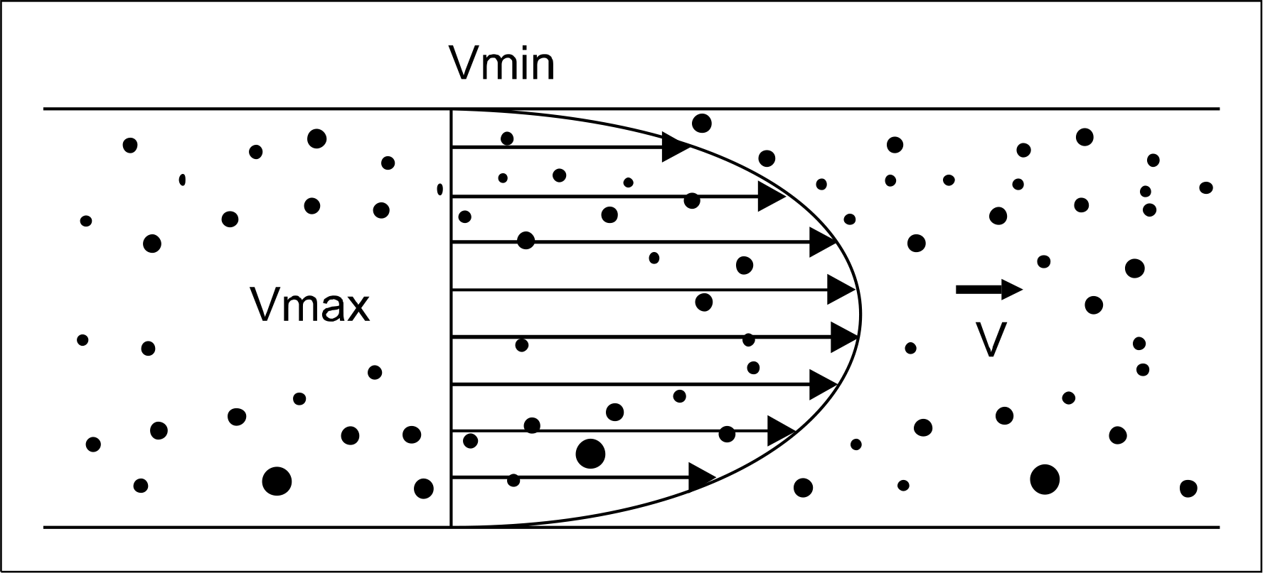 | Fig 3.Blood flow velocity distribution. Vmax, Maximum velocity of blood flow; Vmin, minimum velocity of blood flow. |
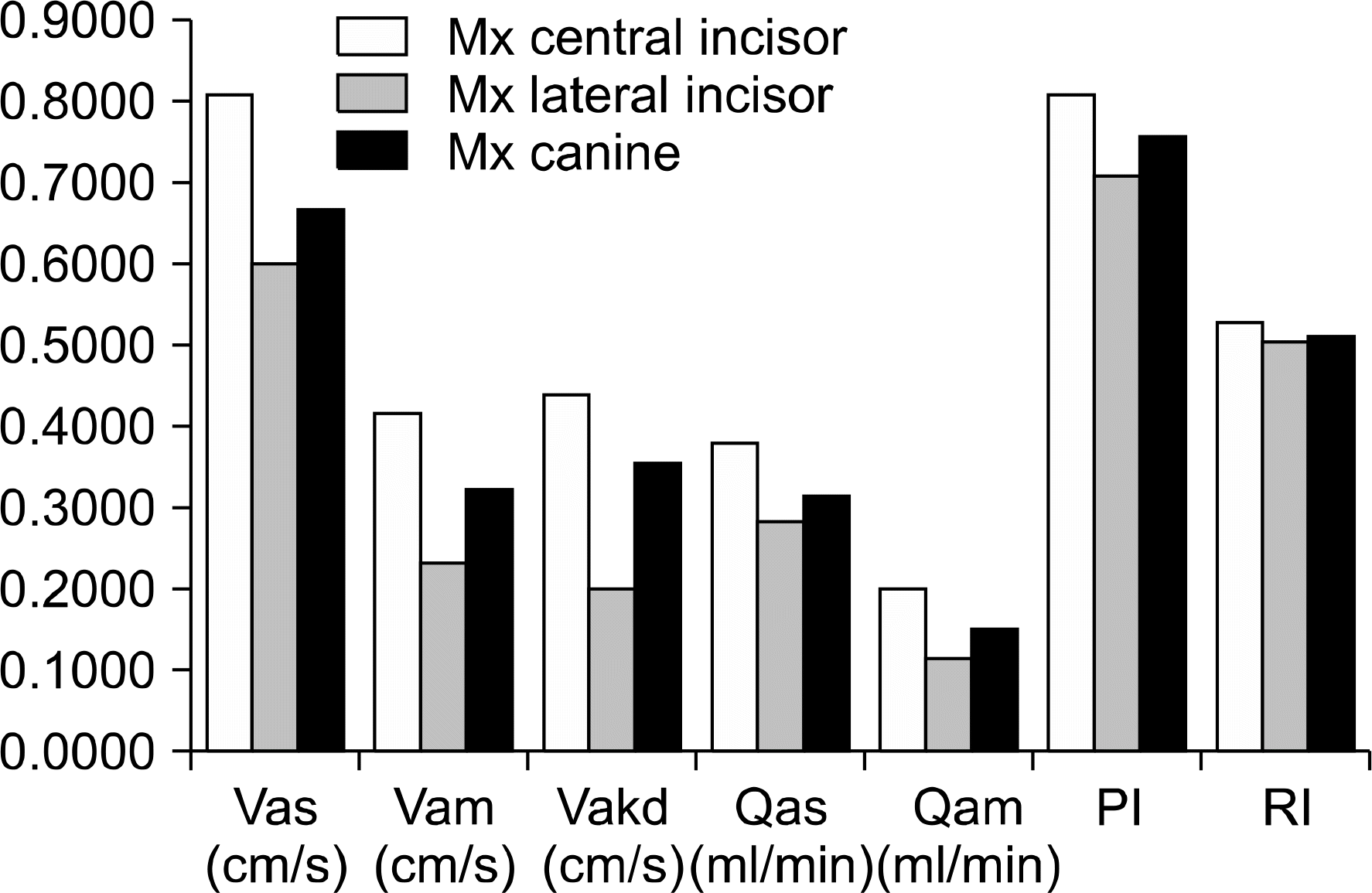 | Fig 4.Comparison of each variable for the maxillary (Mx) teeth before orthodontic treatment. Variable and it’s unit are shown together on horizontal line of graph. |
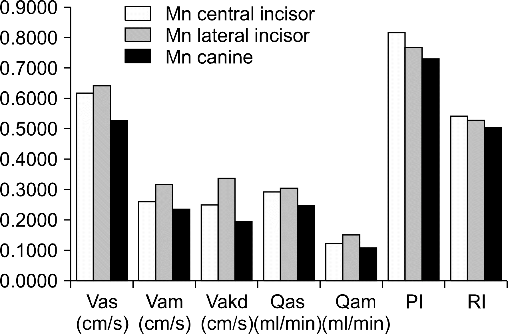 | Fig 5.Comparison of each variable for the mandibular (Mn) teeth before orthodontic treatment. Variable and it’s unit are shown together on horizontal line of graph. |
Table 1.
Definition of Doppler ultrasound variables
Table 2.
Comparison of pulpal blood flow change of 0, 3, 6 weeks
Table 3.
Comparison of pulpal blood flow change of 0, 3, 6 weeks at maxilla (Mx) and mandible (Mn)
Table 4.
Comparison of RI according to position
| Position | Mean | SD | Sig | N |
|---|---|---|---|---|
| Maxilla | 0.52 | 0.07 | * | 27 |
| Mandible | 0.49 | 0.07 | 27 |
Table 5.
Comparison of pulpal blood flow change (Vakd) in maxillary (Mx) central incisor and lateral incisor between 0 week and 6 week
| Tooth | 0 week | 3 weeks | 6 weeks | Sig | N | |||
|---|---|---|---|---|---|---|---|---|
| Mean | SD | Mean | SD | Mean | SD | |||
| Mx central incisor | 0.43 | 0.15 | 0.25 | 0.03 | 0.30 | 0.18 | * | 9 |
| Mx lateral incisor | 0.20 | 0.12 | 0.34 | 0.24 | 0.41 | 0.15 | 9 | |
Table 6.
Comparison of pulpal blood amount on each teeth of Mx. & Mn. before treatment
 XML Download