

 PDF
PDF ePub
ePub Citation
 Print
Print


Abstract
Objective
Miniscrews are widely used in orthodontic treatment for the purpose of anchorage control. Maximum anchorage can be acquired by the use of miniscrews. Maxillary miniscrew has many clinical advantage for orthodontic treatment. Maxillary sinus, tooth root can be an obstacle for maxillary miniscrew installation. The purpose of this study was to find the safest area and direction of miniscrew insertion in consideration of the maxillary sinus.
Methods
The maxillary sinus area of 40 patients (20 male, 20 female) was measured using 3D computed tomography and 3D reconstruction program.
Results
The maxillary sinus floor was located most inferiorly between the 1st molar and 2nd molar and located most superiorly between the 1st premolar and 2nd premolar. Buccal bone thickness from the maxillary sinus is significantly thicker between the 1st molar and 2nd molar and significantly thinner between the 1st premolar and 2nd premolar. The area between the 1st premolar and 2nd premolar has a significantly longer vertical distance from CEJ to sinus in consideration of buccal bone thickness.
Figures and Tables
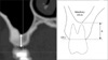
 | Fig. 2Definition of horizontal bone thickness (B) from sinus floor to buccal bone margin in axial view.
|
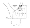 | Fig. 3Vertical bone thickness between CEJ and sinus floor when horizontal bone thickness is 4 mm (C1), 3 mm (C2), and 2 mm (C3) in frontal view.
|
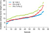 | Fig. 4Available vertical bone height from CEJ when horizontal bone thickness is 4 mm for all 80 samples. Pm, Maxillary premolar; M, maxillary molar.
|


References
1. Misch CE. Contemporary implant dentistry, chapter 8. 1999. 2nd ed. St Louis: Mosby;113.
2. Wehrbein H, Merz BR, Diedrich P. Palatal bone support for orthodontic implant anchorage-a clinical and radiological study. Eur J Orthod. 1999. 21:65–70.

3. Henriksen B, Bavitz B, Kelly B, Harn SD. Evaluation of bone thickness in the anterior hard palate relative to midsagittal orthodontic implants. Int J Oral Maxillofac Implants. 2003. 18:578–581.
4. Bernhart T, Vollgruber A, Gahleitner A, Dörtbudak O, Haas R. Alternative to the median region of the palate for placement of an orthodontic implant. Clin Oral Implants Res. 2000. 11:595–601.
5. Kyung SH. A study on the bone thickness of midpalatal suture area for miniscrew insertion. Korean J Orthod. 2004. 34:63–70.
6. Park YC, Lee JS, Kim DH. Anatomical characteristics of the midpalatal suture area for miniscrew implantation using CT image. Korean J Orthod. 2005. 35:35–42.
7. Gahleitner A, Podesser B, Schick S, Watzek G, Imhof H. Dental CT and orthodontic implants: imaging technique and assessment of available bone volume in the hard palate. Eur J Radiol. 2004. 51:257–262.
8. Lee KY, Lee JW. Study of miniscrew about installation position and condition on maxillary palatal side [thesis]. 2008. Cheonan: Dankook University.
9. Park HS. An anatomical study using CT images for the implantation of micro-implants. Korean J Orthod. 2002. 32:435–441.
10. Lee KJ, Joo E, Kim KD, Lee JS, Park YC, Yu HS. Computed tomographic analysis of tooth-bearing alveolar bone for orthodontic miniscrew placement. Am J Orthod Dentofacial Orthop. 2009. 135:486–494.
11. Jo HS, Lee JW. Three dimensional study of miniscrew about installation area and angle. J Korean Acad Stomatog Func Occl. 2008. 24:203–211.
12. Kim BY, Kim JD. A radiographic study on the morphology of the maxillary sinus. Korean J Oral Maxillofac Radiol. 1991. 21:297–306.
13. Yoon HR, Park CS. A radiologic study of the relationship of the maxillary sinus floor and apex of the maxillary molar. Korean J Oral Maxillofac Radiol. 1998. 28:111–126.
14. Kim SH, Lee KB, Chung KR, Nelson G, Kim TW. Severe bimaxillary protrusion with adult periodontitis treated by corticotomy and compression osteogenesis. Korean J Orthod. 2009. 39:54–65.
15. Lee HA, Park YC. Treatment and posttreatment changes following intrusion of maxillary posterior teeth with miniscrew implants for open bite correction. Korean J Orthod. 2008. 38:31–40.
16. Kim HJ, Yun HS, Park HD, Kim DH, Park YC. Soft-tissue and cortical-bone thickness at orthodontic implant sites. Am J Orthod Dentofacial Orthop. 2006. 130:177–182.
17. Doud Galli SK, Lebowitz RA, Giacchi RJ, Glickman R, Jacobs JB. Chronic sinusitis complicating sinus lift surgery. Am J Rhinol. 2001. 15:181–186.
18. Van den Bergh JP, ten Bruggenkate CM, Disch FJ, Tuinzing DB. Anatomical aspects of sinus floor elevations. Clin Oral Implant Res. 2000. 11:256–265.
19. Vlassis JM, Fugazzotto PA. A classification system for sinus membrane perforations during augmentation procedures with options for repair. J Periodont. 1999. 70:692–699.
20. Pikos MA. Maxillary sinus membrane repair: report of a technique for large perforations. Implant Dent. 1999. 8:29–34.
21. Poggio PM, Incorvati C, Velo S, Carano A. "Safe zones": a guide for miniscrew positioning in the maxillary and mandibular arch. Angle Orthod. 2006. 76:191–197.
22. Choi SY, Lee SR, Koh KJ. A computed tomographic study on the size and bone wall thickness of the maxillary sinus in normal, preoperative and postoperative maxillary sinusitis patients. Korean J Oral Maxillofac Radiol. 2000. 30:109–116.
 XML Download