

 PDF
PDF ePub
ePub Citation
 Print
Print


Abstract
Objective
The purpose of this study was to evaluate the correlation between menton deviation and dental compensation in facial asymmetry.
Methods
Tooth axis and distance of first molar and canine to the reference plane were investigated by cone-beam computerized tomography. The subjects consisted of 50 patients with asymmetric mandibles (male 21, female 29, mean age 24.3 years). Control groups were also assessed (male 11, female 9, mean age 25.6 years). Nine measurements (5 linear measurements and 4 angular measurements) were measured in order to evaluate the correlation between menton deviation and the linear and angular difference of first molar and canine in the deviated and none-deviated sides using the defined MPR images. The differences between deviated and non-deviated side, according to menton deviation, were statistically analyzed using stepwise multiple regression analysis.
Results
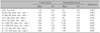
From the result, Menton deviation was negatively correlated with mandibular first molar's angular measurement (Δ∠LM6-Mn plane (dev.-ndev.)) and positively with maxillary fist molar's angular measurement (Δ∠UM6-FH plane (dev.-ndev.)) (p < 0.01). Two angular measurements (Δ∠ LM6-Mn plane (dev.-ndev.), Δ∠UM6-FH plane (dev.-ndev.)) explained the variability in menton deviation with a significant r2 value of 0.589.
Figures and Tables
Fig. 1
Reference planes. A, F-H plane was constructed by connecting both sides of Po and right of Or. MSR plane was perpendicular to FH plane passing through Na and Ba; B, Mn plane was constructed by Me and both sides of Go.
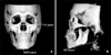
Fig. 2
Linear and angular measurements. A, Distance and angle of U3-FH plane on Ray-sum image which was set by overlapped images on the reference planes; B, distance and angle of UM6-FH plane on MRP image; C, distance and angle of LM6-Mn plane on MRP image.


References
1. Peck H, Peck S. A concept of facial esthetics. Angle Orthod. 1970. 40:284–318.
2. Simon PW. Fundamental principles of a systematic diagnosis of dental anomalies. 1926. Boston: Stratford.
3. Grayson B, Cutting C, Bookstein FL, Kim H, McCarthy JG. The three-dimensional cephalogram: theory, technique, and clinical application. Am J Orthod Dentofacial Orthop. 1988. 94:327–337.

4. Kusnoto B, Evans CA, BeGole EA, de Rijk W. Assessment of 3-dimensional computer-generated cephalomtric measurements. Am J Orthod Dentofacial Orthop. 1999. 116:390–399.
5. Kim WS, Lee KH, Hwang HS. Comparison of asymmetric degree between maxillofacial hard and soft tissue in facial asymmetric subjects using three-dimensional computed tomography. Korean J Orthod. 2005. 35:163–173.
6. Kwon TG, Park HS, Ryoo HM, Lee SH. comparison of craniofacial morphology in patients with and without facial asymmetry--a three-dimensional analysis with computed tomography. Int J Oral Maxillofac Surg. 2006. 35:43–48.
7. Kim GW, Kim JH, Lee KH, Hwang HS. Reproducibility of asymmetry measurements of the mandible in three-dimensional CT imaging. Korean J Orthod. 2008. 38:314–327.
8. Park JW, Kim N, Chang YI. Comparison of landmark position between conventional cephalometric radiography and CT scans projected to midsagittal plane. Korean J Orthod. 2008. 38:427–436.
9. Chang HS, Baik HS. A proposal of landmarks for craniofacial analysis using three-dimensional CT imaging. Korean J Orthod. 2002. 32:313–325.
10. Yoon SJ, Lim HJ, Kang BC, Hwang HS. Three dimensional CT analysis of facial asymmetry. Korean J Oral Maxiillofac Radiol. 2007. 37:45–51.
11. Hinds EC, Reid LC, Burch RJ. Classification and management of mandibular asymmetry. Am J Surg. 1960. 100:825–834.
12. Ahn JS, Hwang HS. Relationship between perception of facial asymmetry and posteroanterior cephalometric measurements. Korean J Orthod. 2001. 31:489–498.
13. Kim GW, Kim JH, Lee KH, Hwang HS. Reproducibility of asymmetry measurements of the mandible in three-dimensional CT imaging. Korean J Orthod. 2008. 38:314–327.
14. Vig PS, Hewitt AB. Asymmetry of the human facial skeleton. Angle Orthod. 1975. 45:125–129.
15. Grayson BH, McCarthy JG, Bookstein F. Analysis of craniofacial asymmetry by multiplane cephalometry. Am J Orthod. 1983. 84:217–224.
16. Fukushima K, Yasui K, Otsuka Y, Matsui S, Hirase N, Takayanagi J, et al. Morphological characteristics of patients with jaw deformity: frontal cephalometric evaluation of facial symmetry. Meikai Univ Dent J. 2003. 32:118–123.
17. Tani M, Iketani M, Watanabe M, Suda S, Fujimura N, Miyazawa M, et al. Posterior-anterior cephalometric analysis in patients with dentofacial deformities. J Jpn Stomatol Soc. 1989. 35:1749–1759.
18. Youn IS, Lee KH, Hwang HS. Clasification of facial asymmetry by cluster analysis. J Korean Dent Assoc. 2001. 39:765–773.
19. Silva MA, Wolf U, Heinicke F, Bumann A, Visser H, Hirsch E. Cone-beam computed tomography for routine orthodontic treatment planning: a radiation dose evaluation. Am J Orthod Dentofaial Orthop. 2008. 133:640e1–e5.
20. Proffit WR. Proffit WR, editor. Concepts of growth and development. Contemporary orthodontics. 2007. 4th ed. St. Louis: Mosby;27–71.
21. Matteson SR, Bechtold W, Phillips C, Staab EV. A method for three-dimensional image reformation for quantitative cephalometric analysis. J Oral Maxillofac Surg. 1989. 47:1053–1061.
22. Proffit WR. Proffit WR, editor. Retention. Contemporary orthodontics. 2007. 4th ed. St. Louis: Mosby;617–631.
 XML Download