

 PDF
PDF ePub
ePub Citation
 Print
Print


Abstract
Objective
The purpose of this study was to investigate the relationship between menarche and cervical vertebral maturation.
Methods
Lateral cephalograms of 67 young korean girls within the range of 1 year before or after their menarche were gathered. The concavity of the cervical vertebrae base and the ratio of the base length to the 3rd and 4th cervical vertebrae anterior height were measured and analyzed.
Results
The mean measured values were as follows; concavity of the 3rd cervical vertebrae base: 1.27 (± 0.18) mm, concavity of the 4th cervical vertebrae base: 1.06 (± 0.15) mm, ratio of the base length to the 3rd cervical vertebrae anterior height: 0.73 (± 0.06) and ratio of the base length to the 4th cervical vertebrae anterior height: 0.70 (± 0.05). There was a significant increase in the ratio of the base length to the 3rd vertebrae anterior height and the base concavity of the 3rd and 4th cervical vertebrae during the period of 1 year before to 1 year after their menarche.
Figures and Tables
Fig 1
Method of measuring the concavity of the cervical vertebrae: distance from the tangent line of the base to the deepest point.
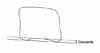
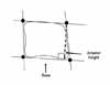
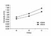
Fig 4
Average curve of the ratio between the length of the base and the anterior height. The same abbreviation as Table 2.
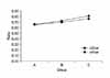
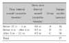
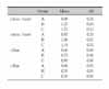
Table 3
Results of statistical comparisons (Kruskal-Wallis test for c3con and c4con, ANOVA with post hoc Scheffe's test for c3bar and c4bar) of measurements for the three groups
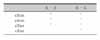
The same abbreviation as Table 2. *p < 0.05.
References
1. Bambha JK, Natta PV. A longitudinal study of occlusion and tooth eruption in relation to skeletal maturation. Am J Orthod. 1959. 45:847–855.

2. Fishman LS. Chronological versus skeletal age, an evaluation of craniofacial growth. Angle Orthod. 1979. 49:181–189.
3. Hägg U, Taranger J. Menarche and voice change as indicators of the pubertal growth spurt. Acta Odontol Scand. 1980. 38:179–186.
4. Hägg U, Taranger J. Maturation indicators and the pubertal growth spurt. Am J Orthod. 1982. 82:299–309.
5. Taranger J, Hägg U. The timing and duration of adolescent growth. Acta Odontol Scand. 1980. 38:57–67.
6. Hägg U, Matsson L. Dental maturity as an indicator of chronological age: the accuracy and precision of three methods. Eur J Orthod. 1985. 7:25–34.
7. Sierra AM. Assessment of dental and skeletal maturity. A new approach. Angle Orthod. 1987. 57:194–208.
8. Coutinho S, Buschang PH, Miranda F. Relationships between mandibular canine calcification stages and skeletal maturity. Am J Orthod Dentofacial Orthop. 1993. 104:262–268.
9. Tanner JM, Whitehouse RH, Marubini E, Resele LF. The adolescent growth spurt of boys and girls of the Harpenden growth study. Ann Hum Biol. 1976. 3:109–126.
10. Green LJ. The interrelationships among height, weight and chronological, dental and skeletal ages. Angle Orthod. 1961. 31:189–193.
11. Grave KC, Brown T. Skeletal ossification and the adolescent growth spurt. Am J Orthod. 1976. 69:611–619.
12. Fishman LS. Radiographic evaluation of skeletal maturation. A clinically oriented method based on hand-wrist films. Angle Orthod. 1982. 52:88–112.
13. Lamparski D. Skeletal age assessment utilizing cervical vertebrae [thesis]. 1972. Pittsburgh: University of Pittsburgh.
14. Hassel B, Farman AG. Skeletal maturation evaluation using cervical vertebrae. Am J Orthod Dentofacial Orthop. 1995. 107:58–66.
15. Mitani H, Sato K. Comparison of mandibular growth with other variables during puberty. Angle Orthod. 1992. 62:217–222.
16. Garcia-Fernandez P, Torre H, Flores L, Rea J. The cervical vertebrae as maturational indicators. J Clin Orthod. 1998. 32:221–225.
17. Bambha JK. Longitudinal cephalometric roentgenographic study of face and cranium in relation to body height. J Am Dent Assoc. 1961. 63:776–799.
18. Bergersen EO. The male adolescent growth spurt: its prediction and relation to skeletal maturation. Angle Orthod. 1972. 42:319–338.
19. Grave KC. Timing of facial growth: a study of relations with stature and ossification in the hand around puberty. Aust Orthod J. 1973. 3:117–122.
20. Hunter CJ. The correlation of facial growth with body height and skeletal maturation at adolescence. Angle Orthod. 1966. 36:44–54.
21. Johnson FE, Hufham HP Jr, Moreschi AF, Terry Gp. Skeletal maturation and cephalofacial development. Angle Orthod. 1965. 35:1–11.
22. Krogman WM. The meaningful interpretation of growth and growth data by the clinician. Am J Orthod. 1958. 44:411–432.
23. Nanda RS. The rates of growth of several facial components measured from serial cephalometric roentgenograms. Am J Orthod. 1955. 41:658–673.
24. Pileski R. Relationship of the ulnar sesamoid and maximum mandibular growth velocity. Am J Orthod. 1973. 43:162–170.
25. Rose GJ. A cross-sectional study of the relationship of facial areas with several body dimensions. Angle Orthod. 1960. 30:6–13.
26. Björk A, Helm S. Prediction of the age of maximum pubertal growth in body height. Angle Orthod. 1967. 37:134–143.
27. Björk A, Skieller V. Facial development and tooth eruption. An implant study at the age of puberty. Am J Orthod. 1972. 62:339–383.
28. Greulich WW, Pyle SI. Radiographic atlas of skeletal development of the hand and wrist. 1959. 2nd ed. Stanford: Stanford University Press.
29. Tanner JM, Whitehouse RH, Cameron N, Marshall WA, Healy MJR, Goldstein H. Assessment of skeletal maturity and prediction of adult height (TW2 method). 1983. 2nd ed. London: Academic Press.
30. Baccetti T, Franchi L, McNamara JA Jr. An improved version of the cervical vertebral maturation (CVM) method for the assessment of mandibular growth. Angle Orthod. 2002. 72:316–323.
31. Lee KH, Hwang YI, Kim YJ, Baek SH, Cha KS, Park YH. Skeletal maturation associated with the fourth cervical vertebra and menarcheal timing. Korean J Orthod. 2008. 38:52–59.
32. Lee CS, Lee SH. A cephalometric study on the ossification pattern of cervical vertebrae in association with skeletal maturity of hand and wrist. J Korean Acad Pediatr Dent. 1992. 19:198–214.
33. Yang KH, Choi NK, Choi BS, Lee YJ, Ryu SY, Kim SM. The skeletal maturity of cervical vertebrae of children with normal occlusion and skeletal class III malocclusion. J Korean Acad Pediatr Dent. 2004. 31:108–113.
34. Demirjian A, Buschang PH, Tanguay R, Patterson DK. Interrelationships among measures of somatic, skeletal, dental, and sexual maturity. Am J Orthod. 1985. 83:433–438.
35. Chang YH, Chung KR. The study on the relationship between the menarche and the bone maturity of malocclusion group. Korean J Orthod. 1995. 25:415–423.
36. Kim KH, Sung SJ, Park SY. Evaluation of the skeletal maturity using the cervical vertebrae and hand-wrist radiographs. Korean J Orthod. 1998. 28:285–295.
37. San Roman P, Palma JC, Oteo MD, Nevado E. Skeletal maturation determined by cervical vertebrae development. Eur J Orthod. 2002. 24:303–311.
 XML Download