

 PDF
PDF ePub
ePub Citation
 Print
Print


Abstract
Objective
The aim of this study was to investigate the changes in the center of resistance of the maxillary teeth in relation to alveolar bone loss.
Methods
A finite element model, which included the upper dentition and periodontal ligament, was designed according to the amount of bone loss (0 mm, 2 mm, 4 mm). The teeth in each group were fixed with buccal and lingual arch wires and splint wires. Retraction and intrusion forces of 200 g for 4 and 6 anterior teeth groups and 400 g for the full dentition group were applied.
Results
The centers of resistance were at 13.5 mm, 14.5 mm, 15 mm apical and 12 mm, 12 mm, 12.5 mm posterior in the 4 incisor group; 13.5 mm, 14.5 mm, 15 mm apical and 14 mm, 14 mm, 14.5 mm posterior in the 6 anterior teeth group; and 11 mm, 13 mm, 14.5 mm apical and 26.5 mm, 27 mm, 25.5 mm posterior in the full dentition group respectively according to 0 mm, 2 mm, 4 mm bone loss.
Figures and Tables
Fig. 1
3-Dimensional finite element mesh of tooth-periodontal ligament (PDL)-alveolar bone of the maxillary dentition. A, Lateral view of maxillary dentition; B, C and D, lateral views of tooth-PDL-alveolar bone model with 0 mm, 2 mm and 4 mm alveolar bone loss respectively.
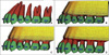
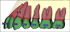
Fig. 2
Finite element models of tooth groups. A, Four anterior teeth; B, six anterior teeth; C, maxillary full dentition. Blue wires on the buccal and palatal surface of the teeth are assumed as rigid body and have no play with brackets, so the movement of individual tooth is limited. Black wires crossing each left and right tooth are designed to distribute the applied force evenly on the dentition; D, vertical and horizontal force application.
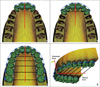
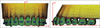
Fig. 4
The vertical position of center of resistance. A, Four anterior teeth: center of resistance (CR) are the points on the Z axis where each bone loss line crosses the sum of displacement (Δy) = 0 line, which are 13.5 mm, 14.5 mm and 15 mm for 0 mm, 2 mm, and 4 mm bone loss respectively; B, six anterior teeth: CR are the points on the Z axis where each bone loss line crosses the sum of displacement (Δy) = 0 line, which are 13.5 mm, 14.5 mm and 15.5 mm for 0 mm, 2 mm, and 4 mm bone loss respectively; C, maxillary full dentition: CR are the points on the Z axis where each bone loss line crosses the sum of displacement (Δy) = 0 line, which are 11 mm, 13 mm and 14.5 mm for 0 mm, 2 mm, and 4 mm bone loss respectively.
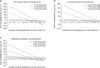
Fig. 5
The horizontal position of center of resistance. A, Four anterior teeth: CR are the points on the Y axis where each bone loss line crosses (or is closest to) the standard deviation of displacement (Δz) = 0 line, which are -12 mm, -12 mm and -12.5 mm for 0 mm, 2 mm, and 4 mm bone loss respectively; B, six anterior teeth: CR are the points on the Y axis where each bone loss line crosses (or is closest to) the standard deviation of displacement (Δz) = 0 line, which are -14 mm, -14 mm and -14.5 mm for 0 mm, 2 mm, and 4 mm bone loss respectively; C, maxillary full dentition: CR are the points on the Y axis where each bone loss line crosses (or is closest to) the standard deviation of displacement (Δz) = 0 line, which are -26.5 mm, -27 mm and -25.5 mm for 0 mm, 2 mm, and 4 mm bone loss respectively.
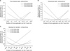
Fig. 6
The vertical and horizontal position of the center of resistance. Round, rectangular, triangle dots indicate the center of resistance of four incisors, six anterior teeth, and maxillary full dentition respectively, and black, blue and green colors represent 0 mm, 2 mm, 4 mm alveolar bone loss respectively.

References
1. Boyd RL, Leggott PJ, Quinn RS, Eakle WS, Chambers D. Periodontal implications of orthodontic treatment in adults with reduced or normal periodontal tissues versus those of adolescents. Am J Orthod Dentofacial Orthop. 1989. 96:191–198.

2. Tanne K, Koenig HA, Burstone CJ. Moment to force ratios and the center of rotation. Am J Orthod Dentofacial Orthop. 1988. 94:426–431.
3. Min YG, Hwang CJ. A study about the change of locations of the center of resistance according to the decrease of alveolar bone heights and root lengths during anterior teeth retraction using the laser reflection technique. Korean J Orthod. 1999. 29:165–181.
4. Smith RJ, Burstone CJ. Mechanics of tooth movement. Am J Orthod. 1984. 85:294–307.
5. Pedersen E, Isidor F, Gjessing P, Andersen K. Location of centres of resistance for maxillary anterior teeth measured on human autopsy material. Eur J Orthod. 1991. 13:452–458.
6. Tanne K, Nagataki T, Inoue Y, Sakuda M, Burstone CJ. Patterns of initial tooth displacements associated with various root lengths and alveolar bone heights. Am J Orthod Dentofacial Orthop. 1991. 100:66–71.
7. Geramy A. Alveolar bone resorption and the center of resistance modification (3-D analysis by means of the finite element method). Am J Orthod Dentofacial Orthop. 2000. 117:399–405.
8. Poppe M, Bourauel C, Jäger A. Determination of the elasticity parameters of the human periodontal ligament and the location of the center of resistance of single-rooted teeth a study of autopsy specimens and their conversion into finite element models. J Orofac Orthop. 2002. 63:358–370.
9. Vollmer D, Bourauel C, Maier K, Jäger A. Determination of the centre of resistance in an upper human canine and idealized tooth model. Eur J Orthod. 1999. 21:633–648.
10. Park GH, Shon BW. The center of resistance of the maxillary anterior segment in the horizontal plane during intrusion by using laser reflection technique. Korean J Orthod. 1993. 23:619–632.
11. Chung KR, Nelson G, Kim SH, Kook YA. Severe bidentoalveolar protrusion treated with orthodontic microimplant-dependent en-masse retraction. Am J Orthod Dentofacial Orthop. 2007. 132:105–115.
12. Park YC, Lee HA, Choi NC, Kim DH. Open bite correction by intrusion of posterior teeth with miniscrews. Angle Orthod. 2008. 78:699–710.
13. Sugawara J, Daimaruya T, Umemori M, Nagasaka H, Takahashi I, Kawamura H, et al. Distal movement of mandibular molars in adult patients with the skeletal anchorage system. Am J Orthod Dentofacial Orthop. 2004. 125:130–138.
14. Coolidge ED. The thickness of the human periodontal membrane. J Am Dent Assoc. 1937. 24:1260–1267.
15. Kronfeld R. Histologic study of the influence of function on the human periodontal membrane. J Am Dent Assoc. 1931. 18:1942.
16. Tanne K, Sakuda M, Burstone CJ. Three-dimensional finite element analysis for stress in the periodontal tissue by orthodontic forces. Am J Orthod Dentofacial Orthop. 1987. 92:499–505.
17. Jeong GM, Sung SJ, Lee KJ, Chun YS, Mo SS. Finite-element investigation of the center of resistance of the maxillary dentition. Korean J Orthod. 2009. 39:83–94.
18. Chung AJ, Kim US, Lee SH, Kang SS, Choi HI, Jo JH, et al. The pattern of movement and stress distribution during retraction of maxillary incisors using a 3-D finite element method. Korean J Orthod. 2007. 37:98–113.
19. Ziegler A, Keilig L, Kawarizadeh A, Jäger A, Bourauel C. Numerical simulation of the biomechanical behaviour of multi-rooted teeth. Eur J Orthod. 2005. 27:333–339.
20. Andrews LF. Straight wire, the concept and appliance. 1989. L.A.: Wells Co..
21. Germane N, Bentley BE Jr, Isaacson RJ. Three biologic variables modifying faciolingual tooth angulation by straight-wire appliances. Am J Orthod Dentofacial Orthop. 1989. 96:312–319.
22. Park CK, Yang WS. A three-dimensional finite element analysis on the location of center of resistance during intrusion of upper anterior teeth. Korean J Orthod. 1997. 27:259–272.
23. Choy K, Kim KH, Burstone CJ. Initial changes of centers of rotation of the anterior segment in response to horizontal forces. Eur J Orthod. 2006. 28:471–474.
24. Vanden Bulcke MM, Burstone CJ, Sachdeva RC, Dermaut LR. Location of the centers of resistance for anterior teeth during retraction using the laser reflection technique. Am J Orthod Dentofacial Orthop. 1987. 91:375–384.
25. Vanden Bulcke MM, Dermaut LR, Sachdeva RC, Burstone CJ. The center of resistance of anterior teeth during intrusion using the laser reflection technique and holographic interferometry. Am J Orthod Dentofacial Orthop. 1986. 90:211–220.
26. Billiet T, de Pauw G, Dermaut L. Location of the centre of resistance of the upper dentition and the nasomaxillary complex. An experimental study. Eur J Orthod. 2001. 23:263–273.
27. Türk T, Elekdag-Türk S, Dinçer M. Clinical evaluation of the centre of resistance of the upper incisors during retraction. Eur J Orthod. 2005. 27:196–201.
28. Yoshida N, Jost-Brinkmann PG, Koga Y, Mimaki N, Kobayashi K. Experimental evaluation of initial tooth displacement, center of resistance, and center of rotation under the influence of an orthodontic force. Am J Orthod Dentofacial Orthop. 2001. 120:190–197.
29. Sia S, Shibazaki T, Koga Y, Yoshida N. Experimental determination of optimal force system required for control of anterior tooth movement in sliding mechanics. Am J Orthod Dentofacial Orthop. 2009. 135:36–41.
30. Choy K, Pae EK, Park Y, Kim KH, Burstone CJ. Effect of root and bone morphology on the stress distribution in the periodontal ligament. Am J Orthod Dentofacial Orthop. 2000. 117:98–105.
31. Ha MH, Son WS. Three-dimensional finite element analysis on intrusion of upper anterior teeth by three-piece base arch appliance according to alveolar bone loss. Korean J Orthod. 2001. 31:209–223.
32. Wakabayashi N, Ona M, Suzuki T, Igarashi Y. Nonlinear finite element analyses: advances and challenges in dental applications. J Dent. 2008. 36:463–471.
33. Cho JH, Park YG, Lee KS. A finite element analysis of the center of resistance of a maxillary first molar. Korean J Orthod. 1993. 23:263–274.
34. Woo JY, Park YC. Experimental study of the vertical location of the centers of resistance for maxillary anterior teeth during retraction using the laser reflection technique. Korean J Orthod. 1993. 23:375–390.
35. Sung SJ, Jeong SJ, Yu YS, Hwang CJ, Pae EK. Customized three-dimensional computational fluid dynamics simulation of the upper airway of obstructive sleep apnea. Angle Orthod. 2006. 76:791–799.
 XML Download