

 PDF
PDF ePub
ePub Citation
 Print
Print


Abstract
Objective
The aim of this study was to find out whether Er:YAG laser can aid in debonding ceramic brackets, and to see what kind of method will be the most appropriate for debonding.
Methods
One hundred and ninety teeth, monocrystalline brackets (MISO™, HT, Ansan-Si, Korea), polycrystalline brackets (Transcend™ series 6000, 3M Untek, Monrovia, CA, USA) and the KEY Laser3 (KavoDental, Biberach, Germany) were used. Experimental groups were classified according to the type of ceramic brackets, and the amount of laser energy (0, 140, 300, 450, 600 mJ). After applying laser on the bracket at two points at 1 pulse each, the shear bond strength was measured. The effect of heat caused by laser was measured at the enamel beneath the bracket and pulp chamber. After measuring the shear bond strength, adhesive residue was evaluated and enamel surface was investigated using SEM.
Results
All ceramic bracket groups showed a significant decrease in shear bond strength as the laser energy increased. The greatest average temperature change was 3.78℃ on the enamel beneath the bracket and 0.9℃ on the pulp chamber. Through SEM, crater shape holes caused by the laser was seen on the enamel and adhesive surfaces.
Figures and Tables
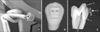 | Fig. 1
A, Laser delivery device with laser focusing guide; B, lasing point (★); C, tooth cross-section. Thermocouple point (T) and lasing point (★). The lasing direction is perpendicular to the slot base.
|
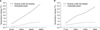 | Fig. 3Temperature change (℃) at various amounts of laser energy on each type of bracket. A, Monocrystalline bracket; B, polycrystalline bracket.
|
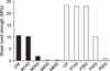
 | Fig. 4Graph of residual adhesive ratings according to the adhesive remnant index (ARI). A, Monocrystalline bracket; B, polycrystalline bracket.
|
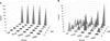
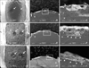 | Fig. 5SEM images of debonding remnant adhesives and tooth of 'Monocrystalline bracket' group. A-C, Group M300; D-F, group M450; G-I, group M600. A and D and G, Remnant adhesive surface on tooth, after laser debonding. Ablated volcano-like hollow was observed (arrow) (original magnification × 30, scale bar: 500 µm); B and E and H, low power SEM image of adhesive-enamel interface, a cross-section view (original magnification × 35, scale bar: 500 µm, e = enamel, r = remnant adhesive); C and F and I, higher magnification of the box shown in B & E & H in cross-sectional view. Ablated volcano-like hollow was also observed. Enamel damage about 10 - 30 µm (▴) (original magnification × 150, scale bar: 100 µm, e = enamel, r = remnant adhesive).
|
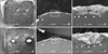 | Fig. 6SEM images of debonding remnant adhesives and tooth of 'Polycrystalline bracket' group. A-C, Group P450; D-F, group P600. A and D, Remnant adhesive surface on tooth, after laser debonding. Ablated volcano-like hollow was observed (arrow). Note that remnant adhesives on the tooth have partially exfoliated (original magnification × 30, scale bar: 500 µm); B and E, low power SEM image of adhesive-enamel interface, a cross-sectional view (original magnification × 35, scale bar: 500 µm, e = enamel, r = remnant adhesive); C and F, higher magnification of the box shown in B & E in cross-sectional view. Ablated volcano-like hollow was also observed. Enamel damage about 10 - 30 µm (▴) (original magnification × 150, scale bar: 100 µm, e = enamel, r = remnant adhesive).
|
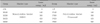
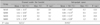
Table 2
Comparison of shear bond strength values (MPa) at various amount of laser energy levels on each type of bracket
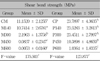
The same abbreviation as Table 1. SD, Standard deviation. *p < 0.001. Entries with the same superscripted letter were not significantly different at p < 0.001.
![]()
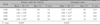
Table 3
Comparison of temperature change (℃) at various amount of laser energy levels on the monocrystalline bracket group
![]()
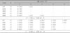
Table 4
Comparison of temperature change (℃) at various amount of laser energy levels on the polycrystalline bracket group
![]()
Table 5
Residual adhesive ratings of the monocrystalline and polycrystalline bracket groups according to the adhesive remnant index (ARI)
*Adhesive remnant index scores were: 1, All adhesive remaining on the tooth; 2, more than 90% of the adhesive remaining on the tooth; 3, more than 10% but less than 90% of the adhesive remaining on the tooth; 4, less than 10% of the adhesive remaining on the tooth; 5, no adhesive remaining on the tooth. a, b: ARI score of group 'CP' was significantly different from group 'P600' (p < 0.05).
![]()
References
1. Bishara SE. Ceramic bracket and the need to develop national standards. Am J Orthod Dentofacial Orthop. 2000. 117:595–597.
2. Arici S, Minors C. The force levels required to mechanically debond ceramic brackets: an in vitro comparative study. Eur J Orthod. 2000. 22:327–334.

3. Bishara SE, Trulove TS. Comparisons of different debonding techniques for ceramic brackets: an in-vitro study. Part 1. Background and methods. Am J Orthod Dentofacial Orthop. 1990. 98:145–153.
4. Storm ER. Debonding ceramic brackets. J Clin Orthod. 1990. 24:91–94.
5. Bishara SE, Trulove TS. Comparisons of different debonding techniques for ceramic brackets: an in-vitro study. Part II. Findings and clinical implications. Am J Orthod Dentofacial Orthop. 1990. 98:263–273.
6. Krell KV, Courey JM, Bishara SE. Orthodontic bracket removal using conventional and ultrasonic debonding techniques, enamel loss, and time requirements. Am J Orthod Dentofacial Orthop. 1993. 103:258–266.
7. Rueggeberg FA, Lockwood P. Thermal debracketing of orthodontic resins. Am J Orthod Dentofacial Orthop. 1990. 98:56–65.
8. Larmour CJ, McCabe JF, Gordon PH. An ex vivo investigation into the effects of chemical solvents on the debond behaviour of ceramic orthodontic brackets. Br J Orthod. 1998. 25:35–39.
9. Strobl K, Bahns TL, Willham L, Bishara SE, Stwalley WC. Laser-aided debonding of orthodontic ceramic brackets. Am J Orthod Dentofacial Orthop. 1992. 101:152–158.
10. Tocchio RM, Willham PT, Mayer FJ, Standing KG. Laser debonding of ceramic orthodontic brackets. Am J Orthod Dentofacial Orthop. 1993. 103:155–162.
11. Mimura H, Deguchi T, Obata A, Yamagishi T, Ito M. Comparison of different bonding materials for laser debonding. Am J Orthod Dentofacial Orthop. 1995. 108:267–273.
12. Rickabaugh JL, Marangoni RD, McCaffrey K. Ceramic bracket debonding with the carbon dioxide laser. Am J Orthod Dentofacial Orthop. 1996. 110:388–393.
13. Ma T, Marangoni RD, Flint W. In vitro comparison of debonding force and intrapulpal temperature changes during ceramic orthodontic bracket removal using a carbon dioxide laser. Am J Orthod Dentofacial Orthop. 1997. 111:203–210.
14. Obata A, Tsumura T, Niwa K, Ashizawa Y, Deguchi T, Ito M. Super pulse CO2 laser for bracket bonding and debonding. Eur J Orthod. 1999. 21:193–198.
15. Kim YJ, Lim SH, Yoon YJ, Park JC, Kim KW. Histologic changes of pulpal tissue after laser-aided ceramic bracket debonding. Korean J Orthod. 2004. 34:343–349.
16. Hayakawa K. Nd:YAG laser for debonding ceramic orthodontic brackets. Am J Orthod Dentofacial Orthop. 2005. 128:638–647.
17. Coluzzi DJ. Fundamentals of dental lasers: science and instruments. Dent Clin North Am. 2004. 48:751–770.
18. Guttenberg SA, Emery RW 3rd. Laser physics and tissue interaction. Oral Maxillofacial Surg Clin North Am. 2004. 16:143–147.
19. Masato M. CO2 laser techniques. 2001. Tokyo: Tohan Co;2–23.
20. Moritz AF, Beer F, Goharkhay K, Schoop U, Strassl M. Oral laser application. 2007. Illinois: Quintessence Pub Co;38–55.
21. International Standards Organization. ISO/TR 106/SC 1/WG 11. Dentistry: Dental materials - Testing of adhesive to tooth structure. 2003.
22. Graber TM, Eliades T, Athanasiou AA. Risk management in orthodontics. 2003. 1st ed. Illinois: Quintessence Pub Co;20–42.
23. Chirila TV, Constable IJ, van Saarloos PP, Barrett GD. Laser-induced damage to transparent polymers: chemical effect of short-pulpsed (Q-switched) Nd:YAG laser radiation on ophthalmic acrylic biomaterials. I. A review. Biomaterials. 1990. 11:305–312.
24. Eliades T, Johnston WM, Eliades G. Direct light transmittance through ceramic brackets. Am J Orthod Dentofacial Orthop. 1995. 107:11–19.
25. Zach L, Cohen G. Pulp response to externally applied heat. Oral Surg Oral Med Oral Pathol. 1965. 19:515–530.
26. Chang JC, Wilder-Smith P. Laser-induced thermal events in empty and pulp-filled dental pulp chambers. Lasers Surg Med. 1998. 22:46–50.
27. Attrill DC, Davies RM, King TA, Dickinson MR, Blinkhorn AS. Thermal effects of the Er:YAG laser on a simulated dental pulp: a quantitative evaluation of the effects of a water spray. J Dent. 2004. 32:35–40.
28. Proffit WR, Fields HW, Sarver DM. Contemporary orthodontics. 2007. 4th ed. St Luis: Mosby Co;415.
29. Delfino CS, Souza-Zaroni WC, Corona SAM, Pécora JD, Palma-Dibb RG. Effect of Er:YAG laser energy on the morphology of enamel/adhesive system interface. Applied Surface Science. 2006. 252:8476–8481.
30. Pus MD, Way DC. Enamel loss due to orthodontic bonding with filled and unfilled resins using various clean-up techniques. Am J Orthod. 1980. 77:269–283.
31. Al Shamsi AH, Cunningham LJ, Lamey PJ, Lynch E. Three-dimensional measurement of residual adhesive and enamel loss on teeth after debonding of orthodontic brackets: an in-vitro study. Am J Orthod Dentofacial Orthop. 2007. 131:301.e9–301.e15.
 XML Download