

 PDF
PDF ePub
ePub Citation
 Print
Print


Abstract
Tooth anklylosis is defined as the adhesion state of alveolar bone to dentin or cementum. Trauma, disturbed metabolic disease, and congenital disease have been given as etiologic factors. Complications of tooth ankylosis are tipping of the neighboring teeth, space loss, and supraeruption of the opposing teeth. Particularly if dental ankylosis occurs in maxillary incisors of a growing child, the ankylosed tooth can not move vertically with subsequent disturbance in vertical growth of the alveolar process. With an appropriate treatment approach, an esthetic condition must be achieved especially in the maxillary anterior region. In this report, two cases are presented which were treated by the surgical repositioning method. One is treated by alveolar bone distraction osteogenesis which used a tooth-borne type distraction device and the other by single tooth osteotomy.
Figures and Tables
Fig. 1
Facial and intraoral photographs and panoramic, standard and cephalometric radiographs before treatment. Maxillary right first premolar, canine, lateral and central incisors showed infraocclusion.
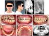
Fig. 2
Tooth separation test for differential diagnosis of tooth ankylosis using elastic module. A and B, Tight contact existed between maxillary right central, lateral incisors and canine; C and D, central incisor was separated; E and F, lateral incisor was separated.
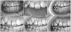
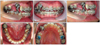

Fig. 5
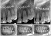
Latency period and distraction procedure. A, Latency period for 5 days; B, 1 mm/day for 4 days; C, D, 0.5 mm/day for 8 days.
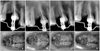
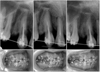

Fig. 8
Facial and intraoral photographs and panoramic, standard and cephalometric radiographs after orthodontic treatment.
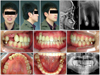
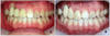
Fig. 10
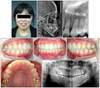
Facial and intraoral photographs and panoramic, occlusal and cephalometric radiographs before treatment. Maxillary left canine was impacted.
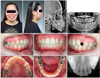
Fig. 11
Progress of orthodontic traction of impacted canine using cantilever spring and indirect anchorage. Orthodontic micro-implant anchorage was splinted to the first molar by 0.019" × 0.025" SS wire. A, Inserting cantilever spring; B, after 1 month; C, after 4 months.
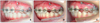

Fig. 13
Surgical repositioning of upper left canine by single tooth osteotomy. A, Flap design; B, incision; C, flap elevation & bone contouring; D, E, fixation by miniplate.

References
1. The Korean Council of the Faculty of Orthodontics. Textbook of orthodontics. 2006. Seoul: Daehannarae Publishing;150.
2. Frazier-Bowers SA, Koehler KE, Ackerman JL, Proffit WR. Primary failure of eruption: Further characterization of a rare eruption disorder. Am J Orthod Dentofacial Orthop. 2007. 131:e1–e11.

3. Andreasen JO. Periodontal healing after replantation and autotransplantation of incisors in monkeys. Int J Oral Surg. 1981. 10:54–61.
4. Biederman W. Etiology and treatment of tooth ankylosis. Am J Orthod. 1962. 48:670–684.
5. Kurol J, Thilander B. Infraocclusion of primary molars and the effect on occlusal development, a longitudinal study. Eur J Orthod. 1984. 6:277–293.
6. Alcan T. A miniature tooth-borne distractor for the alignment of ankylosed teeth. Angle Orthod. 2006. 76:77–83.
7. Kofod T, Würtz V, Melsen B. Treatment of an ankylosed central incisor by single tooth dento-osseous osteotomy and a simple distraction device. Am J Orthod Dentofacial Orthop. 2005. 127:72–80.
8. Razdolsky Y, El-Bialy TH, Dessner S, Buhler JE Jr. Movement of ankylosed permanent teeth with a distraction device. J Clin Orthod. 2004. 38:612–620.
9. Kinzinger GS, Jänicke S, Riediger D, Diedrich PR. Orthodontic fine adjustment after vertical callus distraction of an ankylosed incisor using the floating bone concept. Am J Orthod Dentofacial Orthop. 2003. 124:582–590.
10. Son WS, Chung IK, Shin SH. Surgically assisted orthodontic treatment of ankylosed maxillary incisor. Korean J Orthod. 2002. 32:257–264.
11. Ilizarov GA. Clinical application of the tension-stress effect for limb lengthening. Clin Orthop Relat Res. 1990. 250:8–26.
12. McCarthy JG, Stelnicki EJ, Grayson BH. Distraction osteogenesis of the mandible: a ten-year experience. Semin Orthod. 1999. 5:3–8.
13. Toth BA, Kim JW, Chin M, Cedars M. Distraction osteogenesis and its application to the midface and bony orbit in craniosynostosis syndromes. J Cranofac Surg. 1998. 9:100–113.
14. Samchukov ML, Cope JB, Cherkashin AM. Craniofacial distraction osteogenesis. 2001. St. Louis: Mosby;379–458.
15. Albers DD. Ankylosis of teeth in the developing dentition. Quintessence Int. 1986. 17:303–308.
16. Hammarström L, Pierce A, Blomlöf L, Feiglin B, Lindskog S. Tooth avulsion and replantation - a review. Endod Dent Traumatol. 1986. 2:1–8.
17. Lindskog S, Pierce AM, Blomlof L, Hammarstrom L. The role of the necrotic periodontal membrane in cementum resorption and ankylosis. Endod Dent Traumatol. 1985. 1:96–101.
18. Malmgren B, Malmgren O. Rate of infraposition of reimplanted ankylosed incisors related to age and growth in children and adolescents. Dent Traumatol. 2002. 18:28–36.
 XML Download