

 PDF
PDF ePub
ePub Citation
 Print
Print


Abstract
Objective
This study investigated the onset, peak height velocity (PHV) and end of adolescent growth spurt as well as menarcheal timing deduced from surveying accumulative height growth over many years.
Methods
Ninety six students of Samgoe high school between 1st and 3rd grade that were in good health participated in the research. A survey questionnaire was distributed to examine the students' health status and menarche timing.
Results
Adolescent growth spurt typically began at the age of 9.9 and reached a PHV at the age of 11.6. The growth spurt ended at the age of 14.1 on average. The average age of menarche was 12.6 years, which was about one year after the PHV of adolescent growth spurt. In most cases, menarche came after PHV. But in 24% of the students, menarche and PHV was nearly coincident or menarche preceded PHV. The growth curves were classified into four types. A typical adolescent growth spurt showed one PHV on graph that drastically drops after the PHV. However, there were cases with two PHVs.
Figures and Tables
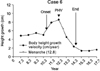
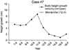
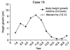
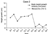
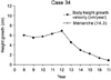
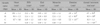
Table 1
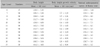
Mean, standard deviation of body height and body height growth velocity at specified ages and comparison with the mean of specified age of National anthropometric survey of Korea of 1997
![]()

References
1. Fishman LS. Chronologic age versus skeletal age, an evaluation of craniofacial growth. Angle Orthod. 1979. 49:181–189.
2. Grave KC, Brown T. Skeletal ossification and the adolescent growth spurt. Am J Orthod. 1976. 69:611–619.

3. Pileski RC, Woodside DG, James GA. Relationship of the ulnar sesamoid bone and maximum mandibular growth velocity. Angle Orthod. 1973. 43:162–170.
4. Hägg U, Taranger J. Skeletal stages of the hand and wrist as indicators of the pubertal growth spurt. Acta Odontol Scand. 1980. 38:187–200.
5. Deicke M, Pancherz H. Is radius-union an indicator for completed facial growth? Angle Orthod. 2005. 75:295–299.
6. Chang YH, Chung KR. The study on the relationship between the menarche and the bone maturity of malocclusion group. Korean J Orthod. 1995. 25:415–423.
7. Kim KH, Baik HS, Son ES. A study on menarche and skeletal maturity among various malocclusion groups. Korean J Orthod. 1998. 28:581–589.
8. Kim KH, Choy KC, Jung KY. The relationship between menarche and the ossification stages of the phalanx of the first and third finger. Korean J Orthod. 2002. 32:265–274.
9. Lamparski DG. Skeletal age assessment utilizing cervical vertebrae. 1972. Pittsburgh: University of Pittsburgh;[Master's thesis].
10. Lee KH, Hwang YI, Kim YJ, Baek SH, Cha KS, Park YH. Skeletal maturation associated with the fourth cervical vertebra and menarcheal timing. Korean J Orthod. 2008. 38:52–59.
11. Lee JH, Kang YG, Lee KS, Nam JH. Maturation of cervical verterae in relation to menarche. Korean J Orthod. 2009. 39:28–35.
12. Tanner JM, Whitehouse RH, Marshall WA, Healy MJ, Goldstein H. Assessment of skeletal maturity and prediction of adult height (TW2 method). 1975. New York: Academic Press.
13. Fishman LS. Maturational patterns and prediction during adolescence. Angle Orthod. 1987. 57:178–193.
14. Demirjian A, Buschang PH, Tanguay R, Patterson DK. Interrelationships among measures of somatic, skeletal, dental and sexual maturity. Am J Orthod. 1985. 88:433–438.
15. Björk A, Helm S. Prediction of age of the maximum pubertal growth in body height. Angle Orthod. 1967. 37:134–143.
16. Bergersen EO. The male adolescent growth spurt: its prediction and relation to skeletal maturation. Angle Orthod. 1972. 42:319–338.
17. Bambha JK. Longitudinal cephalometric roentgenographic study of face and cranium in relation to body height. J Am Dent Assoc. 1961. 63:776–799.
18. Hägg U, Taranger J. Maturation indicators and the pubertal growth spurt. Am J Orthod. 1982. 82:299–309.
19. Korean agency for technology and standards. The report of national anthropometric survey of Korea. 1997.
20. Riley AP, Samuelson JL, Huffman SL. Gray R, editor. The relationship of age at menarche and fertility in undernourished adolescents. Biomedical and demographic determinants of reproduction. 1993. Oxford: Clarendon Press.
21. Adair LS. Size at birth predicts age at menarche. Pediatrics. 2001. 107:E59.
22. Eveleth PB, Tanner JM. Worldwide variation in human growth. 1991. Cambridge: Cambridge University Press.
23. Kaplowitz PB, Slora EJ, Wasserman RC, Pedlow SE, Herman-Giddens ME. Earlier onset of puberty in girls: relation to increased body mass index and race. Pediatrics. 2001. 108:347–353.
24. Hwang JY, Shin C, Frongillo EA, Shin KR, Jo I. Secular trend in age at menarche for South Korean women born between 1920 and 1986: the Ansan study. Ann Hum Biol. 2003. 30:434–442.
25. Bagga A, Kulkarni S. Age at menarche and secular trend in Maharashtrian (Indian) girls. Acta Biologica Szegediensis. 2000. 44:53–57.
26. Kac G, Auxiliadora de Santa Cruz Coel, Velasquez-Melendez G. Secular trend in age at menarche for women born between 1920 and 1979 in Rio de Janeiro, Brazil. Ann Hum Biol. 2000. 27:423–428.
27. Chumlea WC, Schubert CM, Roche AF, Kulin HE, Lee PA, Himes JH, et al. Age at menarche and racial comparisons in US girls. Pediatrics. 2003. 111:110–113.
28. Wu T, Mendola P, Buck GM. Ethnic differences in the presence of secondary sex characteristics and menarche among US girls: the Third National Health and Nutrition Examination Survey, 1988-1994. Pediatrics. 2003. 110:752–757.
29. Lee SJ, Chung KR, Park YG. The study of the changes in skeletal maturity according to the time passed from menarche. Korean J Orthod. 1998. 28:409–417.
30. Zacharias L, Rand WM. Adolescent growth in height and its relation to menarche in contemporary American girls. Ann Hum Biol. 1983. 10:209–222.
31. Abbassi V. Growth and normal puberty. Pediatrics. 1998. 102:507–511.
32. Tanner JM, Davies PS. Clinical longitudinal standards for height and height velocity for North American children. J Pediatr. 1985. 107:317–329.
33. Sizonenko PC. Normal sexual maturation. Pediatrician. 1987. 14:191–201.
34. Hägg U, Taranger J. Menarche and voice change as indicators of the pubertal growth spurts. Acta Odontol Scand. 1980. 38:179–186.
35. Gasser T, Müller HG, Köhler W, Prader A, Largo R, Molinari L. An analysis of the mid-growth and adolescent spurts of height based on acceleration. Ann Human Biol. 1985. 12:129–148.
36. Nanda RS. The rates of growth of several facial components measures from serial cephalometric roentgenograms. Am J Orthod. 1955. 41:658–673.
37. Thompson GW, Popovich F. Relationship of craniofacial changes and skeletal age increments in females. Hum Biol. 1973. 45:595–603.
 XML Download