

 PDF
PDF ePub
ePub Citation
 Print
Print


Abstract
Objective
The purpose of this study was to reveal the position of the incisive foramen in relation to the incisive papilla and cusp tips.
Methods
Plaster models and CT images of 25 adult orthodontic patients were used to measure the width of the incisive canal and positions of the anterior and posterior borders of the incisive foramen in relation to the incisive papilla.
Results
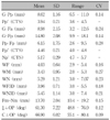
The palatal surface distance from the interdental papilla between the maxillary central incisors to the posterior border of the incisive foramen along the palatal surface was 1.7 fold of the distance from the interdental papilla between the central incisors to the posterior border of the incisive papilla. The distance between the posterior border of the incisive papilla and posterior border of the incisive foramen along the palatal surface was 6.15 ± 1.75 mm. The anteroposterior position of the posterior border of the incisive foramen was slightly anterior to the lingual cusp tips of the maxillary 1st premolars. The width of the incisive foramen was 4.03 ± 0.64 mm, therefore it is recommended to position the mini-implant more than 3 mm laterally when placing a mini-implant lateral to the incisive foramen, from the center.
Figures and Tables
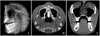 | Fig. 1Adjustment of reference lines. A, Occlusal plane in sagittal section - from incisal edge of maxillary central incisor to mesiobuccal cusp tip of maxillary 1st molar; B, anterior posterior line in axial section - from ANS to PNS; C, transverse line in coronal section - a line connecting lingual cusp tips of maxillary 1st molars.
|
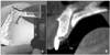
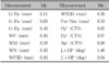
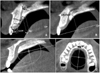 | Fig. 2Landmarks and measurements made in the midsagittal sections. A, Landmarks, linear and angular mearsuremnets in midsagittal plane; B, measurement of bone thickness at the cusp tip areas. ⊥Surface, measurement along the perpendicular line to the tangent line to the bone surface; ⊥OP, measurement along the perpendicular line to the occlusal plane; C, mesiodistal width of incisive canal on axial section. Refer to Table 1.
|
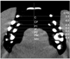 | Fig. 3CTS (Cusp Tip Scale: anteroposterior numerical scale by cusp tip) was used to evaluate anteroposterior position of posterior border of incisive foramen and palatal bone thickness at each cusp tip area. C, Canine cusp tip scored as 3.5; CP, midpoint between canine cusp tip and lingual cusp tip of 1st premolar scored as 4.0; P1, lingual cusp tip of 1st premolar scored as 4.5; IP, midpoint between lingual cusp tips of 1st and 2nd premolars scored as 5.0; P2, lingual cusp tip of 2nd premolar scored as 5.5; PM, midpoint between lingual cusp tips of 2nd premolar and mesiolingual cusp tip of 1st molar scored as 6.0; Mm, mesioligual cusp tip of 1st molar scored as 6.5.
|
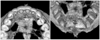 | Fig. 4Mesiodistal width of incisive foramen on 3-D view. WF3D, Width of incisive foramen of palate; WN3D, width of nasal aperture of incisive canal at nasal cavity.
|
 | Fig. 5
A, G-Pp length measurement on plaster model; B, cusp tip scale of posterior border of papilla on midsagittal section. Refer to Table 1.
|
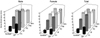 | Fig. 6Midpalatal bone thickness at cusp tip areas. P1, Lingual cusp tip of 1st premolar; IP, midpoint between lingual cusp tips of 1st and 2nd premolars; P2, lingual cusp tip of 2nd premolar; PM, midpoint between lingual cusp tip of 2nd premolar and mesiolingual cusp tip of 1st molar; Mm, mesioligual cusp tip of 1st molar; OP, occlusal plane.
|
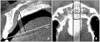 | Fig. 7Canal zone. A, Sagittal section. Anterior border of canal zone is on the perpendicular line to occlusal plane starting from the posterior border of incisive foramen. Posterior border of canal zone is on the perpendicular line to the occlusal plane starting from the posterior border of nasal aperture of incisive canal; B, Axial section. Lateral border of canal zone is along the lateral border of incisive canal.
|
References
1. Lee HA, Park YC. Treatment and posttreatment changes following intrusion of maxillary posterior teeth with miniscrew implants for open bite correction. Korean J Orthod. 2008. 38:31–40.

2. Kim SH, Lee KB, Chung KR, Nelson G, Kim TW. Severe bimaxillary protrusion with adult periodontitis treated by corticotomy and compression osteogenesis. Korean J Orthod. 2009. 39:54–65.
3. Kyung SH. A study on the bone thickness of midpalatal suture area for miniscrew insertion. Korean J Orthod. 2004. 34:63–70.
4. Park YC, Lee JS, Kim DH. Anatomical characteristics of the midpalatal suture area for miniscrew implantation using CT image. Korean J Orthod. 2005. 35:35–42.
5. Kraut RA, Boyden DK. Location of incisive canal in relation to central incisor implants. Implant Dent. 1998. 7:221–225.
6. Cavalcanti MG, Yang J, Ruprecht A, Vannire MW. Accurate linear measurements in the anterior maxilla using orthoradially reformatted sprial computed tomography. Dentomaxillofac Radiol. 1999. 28:137–140.
7. Kim GT, Hwang EH, Lee SR. A study of incisive canal using a cone beam computed tomography. Korean J Oral Maxillofac Radiol. 2004. 34:7–12.
8. Henriksen B, Bavitz B, Kelly B, Harm SD. Evaluation of bone thickness in the anterior palate relative to midsagittal orthodontic implants. Int J Oral Maxillofac Implants. 2003. 18:578–581.
9. Mraiwa N, Jacobs R, Van Cleynenbreugel J, Sanderink G, Schutyser F, Suetens P, et al. The nasopalatine canal revisited using 2D and 3D CT imaging. Dentomaxillofac Radiol. 2004. 33:396–402.
10. Wehrbein H, Merz BR, Diedrich P. Palatal bone support for orthodontic implant anchorage - a clinical and radiological study. Eur J Orthod. 1999. 21:65–70.
11. Bernhart T, Vollgruber A, Gahleitner A, Dörtbudak O, Haas R. Alternative to the median region of the palate for placement of an orthodontic implant. Clin Oral Implants Res. 2000. 11:595–601.
12. Kang S, Lee SJ, Ahn SJ, Heo MS, Kim TW. Bone thickness of the palate for orthodontic mini-implant anchorage in adults. Am J Orthod Dentofacial Orthop. 2007. 131:Suppl. S74–S81.
13. King KS, Lam EW, Faulkner MG, Heo G, Major PW. Vertical bone volume in the paramedian palate of adolescents: a computed tomography study. Am J Orthod Dentofacial Orthop. 2007. 132:783–788.
14. Ferner H, Staubesand J. Sobotta atlas of human anatomy Vol. I: head, neck, upper extrimities. 1983. Seoul: Il-Joong Sa;10.
15. Houston WJ. The analysis of errors in orthodontic measurements. Am J Orthod. 1983. 83:382–390.
16. Orban BJ. Oral histology and embryology. 1953. 3rd ed. St. Louis: The CV Mosby Co;13–28.
17. Patten BM. Human embryology. 1946. New York: The Blakiston Co;284427–434.
18. Sicher H, DuBrul EL. Oral anatomy. 1970. 5th ed. St. Louis: The CV Mosby Co;37–38. 68–69.
19. Langford RJ. The contrubution of the nasopalatine nerve to sensation of the hard palate. Br J Oral Maxillofac Surg. 1989. 27:379–386.
20. Burdi AR. Distribution of midpalatal cysts: a reevaluation of human palatal closure mechanisms. J Oral Surg. 1968. 26:41–45.
21. Zingeser MR. The nasopalatine ducts and associated structures in the rhesus monkey (Macaca mulatta): topography, prenatal development, function, and phylogeny. Am J Anat. 1984. 170:581–595.
22. Van der Waal I, Van der Kwast WAM. Oral pathology. 1988. Chicago: Quintessence Publishing Co;145–146.
23. Jacob S, Zelano B, Gungor A, Abbott D, Naclerio R, McClintock MK. Location and gross morphology of the nasopalatine duct in human adults. Arch Otolaryngol Head Neck Surg. 2000. 126:741–748.
24. Kim MK. Head and neck applied anatomy. 1985. Seoul: Eu-chi-hak Sa;15.
25. Bodin I, Isacsson G, Julin P. Cysts of the nasopalatine duct. Int J Oral Maxillofac Surg. 1986. 15:696–706.
26. Triaca A, Antonini M, Wintermantel E. Ein neues Titan-Flanchschrauben-Implantat zur orthodontischen Verankerung am anterioren Gaumen. Informationen aus der orthodontischen Kieferorthopadie. 1992. 24:251–257.
27. Block MS, Hoffman DR. A new device for absolute anchorage for orthodontics. Am J Orthod Dentofacial Orthoped. 1995. 107:251–258.
28. Wehrbein H, Merz BR, Diedrich P, Glatzmaier J. The use of palatal implants for orthodontic anchorage. Design and clinical application of the orthosystem. Clin Oral Implants Res. 1996. 7:410–416.
29. Wehrbein H, Merz BR. Aspects of the use of endosseous palatal implants in orthodontic therapy. J Esthetic Dent. 1998. 10:315–324.
30. Tosun T, Keles A, Erverdi N. Method for the placement of palatal implants. Int J Oral Maxillofac Implants. 2002. 17:95–100.
31. Park YC, Kim JG, Lee JS. Atlas of contemparary orthodontics. 2006. Vol. III:Shin-Hueng International;168.
32. Wehrbein H, Yildizhan F. The midpalatal suture in young adults. A radiological-histological investigation. Eur J Orthod. 2001. 23:105–114.
33. Schlegel KA, Kinner F, Schlegel KD. The anatomic basis for palatal implants in orthodontics. Int J Adult Orthod Orthognath Surg. 2002. 17:133–139.
34. Persson M, Thilander B. Palatal suture closure in man from 15 to 35 years of age. Am J Orthod. 1977. 72:42–45.
35. Latham RA. The structure and development of the intermaxillary suture. J Anat. 1970. 106:167.
36. Kim HJ, Yun HS, Park HD, Kim DH, Park YC. Soft-tissue and cortical bone thickness at orthodontic implant sites. Am J Orthod Dentofacial Orthop. 2006. 130:177–182.
 XML Download