

 PDF
PDF ePub
ePub Citation
 Print
Print


Abstract
Objective
CBCT has become popular for orthodontic diagnosis and treatment planning in recent times. The 3D pharyngeal airway space needs to be analysed using a 3D diagnostic tool. The aim of this study was to analyse the pharyngeal airway of different craniofacial morphology using CBCT.
Methods
The sample compromised 102 subjects divided into 3 groups (Class I, II, III) and 6 subgroups according to normal or vertical craniofacial patterns. All samples had CBCT (VCT, Vatech, Seoul, Korea) taken for orthodontic treatment. The pharyngeal airway was assessed according to the reference planes: aa plane (the most anterior point on the anterior arch of atlas), CV2 plane, and CV3 plane (most infero-anterior point on the body of the second & third cervical vertebra). The intergroup comparison was performed with one-way ANOVA and duncan test as a second step.
Results
The results showed the pharyngeal airway and anteroposterior width of group 2 (Class II) in aa plane, CV2 plane, CV3 plane were significant narrower than in group 3 (Class III). There was no significant difference between vertical and normal craniofacial patterns except for the anteroposterior pharyngeal width of Group 1 (Class I) in aa plane.
Figures and Tables
Fig. 1
A, Reference plane (FH plane, aa plane, CV2 plane, CV3 plane); B, measurements of pharyngeal airway width and area on the reference plane.
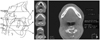
Fig. 2
A, Pharyngeal airway areas of subgroups (vertical and normal pattern) in aa plane; B, pharyngeal airway areas of subgroups (vertical and normal pattern) in CV2 plane; C, pharyngeal airway areas of subgroups (vertical and normal pattern) in CV3 plane.
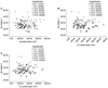
Fig. 3
A, Pharyngeal airway width (apw) of subgroups (vertical and normal pattern) in aa plane; B, pharyngeal airway width (apw) of subgroups (vertical and normal pattern) in CV3 plane.
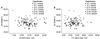
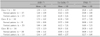

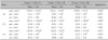
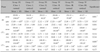
Table 4
Means and standard deviations of area, anteroposterior width (apw) and transverse width (tsw) and results of ANOVA, followed by Duncan test
References
1. Moss ML, Salentijn L. The primary role of functional matrices in facial growth. Am J Orthod. 1969. 55:566–577.

2. Dunn GF, Green LJ, Cunat JJ. Relationship between variation of mandibular morphology and variation of nasopharyngeal airway size in monozygotic twins. Angle Orthod. 1973. 43:129–135.
3. Kerr WJ. The nasopharynx, face height, and overbite. Angle Orthod. 1985. 55:31–36.
4. McNamara JA Jr. A method of cephalometric evaluation. Am J Orthod. 1984. 86:449–469.
5. Hwang YI, Lee KH, Lee KJ, Kim SC, Cho HJ, Cheon SH, et al. Effect of airway and tongue in facial morphology of prepubertal Class I, II children. Korean J Orthod. 2008. 38:74–82.
6. Lee YS, Kim JC. A cephalometric study on the airway size according to the types of the malocclusion. Korean J Orthod. 1995. 25:19–20.
7. Son WS, Choi YS. Evaluation of hyoid bone position and airway size in Class III malocclusion. Korean J Orthod. 1996. 26:247–254.
8. Kwak SY, Kim HY, Jeon YM, Kim JG. Airway size in mlocclusions with hyperdivergent skeletal pattern. Korean J Orthod. 2003. 33:293–305.
9. Kyung SH, Park YC, Pae EK. Obstructive sleep apnea patients with the oral appliance experience pharyngeal size and shape changes in three dimensions. Angle Orthod. 2005. 75:15–22.
10. Rachmiel A, Aizenbud D, Pillar G, Srouji S, Peled M. Bilateral mandibular distraction for patients with compromised airway analyzed by three-dimensional CT. Int J Oral Maxillofac Surg. 2005. 34:9–18.
11. Kim HJ, Park HS, Kwon OW. Evaluation of potency of panoramic radiography for estimating the position of maxillary impacted canines using 3D CT. Korean J Orthod. 2008. 38:265–274.
12. Jeon HJ, Park SH, Jung SH, Chun YS. Three dimensional analysis of tooth movement using different sizes of NiTi wire on NiTi scissors-bite corrector. Korean J Orthod. 2009. 39:43–53.
13. Hwang SH, Park IS, Nam KY, Kim JB, Cho YW, Suh YS, et al. Cephalometric differences in obstructive sleep apnea between obese and non-obese Korean male patients. Korean J Orthod. 2008. 38:202–213.
14. Major MP, Flores-Mir C, Major PW. Assessment of lateral cephalometric diagnosis of adenoid hypertrophy and posterior upper airway obstruction: a systemic review. Am J Orthod Dentofaca Orthop. 2006. 130:700–708.
15. Jeans WD, Fernando DC, Maw AR, Leighton BC. A longitudinal study of the growth of the nasopharynx and its contents in normal children. Br J Radiol. 1981. 54:117–121.
16. Lagravère MO, Carey J, Toogood RW, Major PW. Three-dimensional accuracy of measurements made with software on cone-beam computed tomography images. Am J Orthod Dentofacial Orthop. 2008. 134:112–116.
17. Chang HS, Baik HS. A proposal of landmarks for craniofacial analysis using three-dimensional CT imaging. Korean J Orthod. 2002. 32:313–325.
18. Ludlow JB, Davies-Ludlow LE, Brooks SL, Howerton WB. Dosimetry of 3 CBCT devices for oral and maxillofacial radiology: CB Mercurary, NewTom 3G and i-CAT. Dentomaxillofac Radiol. 2006. 35:219–226.
19. Danforth RA, Dus I, Mah J. 3-D volume imaging for dentistry: a new dimension. J Calf Dent Assoc. 2003. 31:817–823.
20. Anegawa E, Tsuyama H, Kusakawa J. Lateral cephalometric analysis of the pharyngeal airway space affected by head posture. Int J Oral Maxillofac Surg. 2008. 37:805–809.
21. de Freitas MR, Alcazar NM, Janson G, de Freitas KM, Henriques JF. Upper and lower pharyngeal airway in subjects with Class I and Class II malocclusions and different growth patterns. Am J Orthod Dentofacial Orthop. 2006. 130:742–745.
22. Ceylan I, Okatay H. A study on the pharyngeal size in different skeletal patterns. Am J Orthod Dentofac Orthop. 1995. 108:69–75.
23. Chen F, Terada K, Hua Y, Saito I. Effects of bimaxillary surgery and mandibular setback surgery on pharyngeal airway measurements in patients with Class III skeletal deformities. Am J Orthod Dentofacial Orthop. 2007. 131:372–377.
 XML Download