

 PDF
PDF ePub
ePub Citation
 Print
Print


INTRODUCTION
Assessing maturational status, regardless of whether the pubertal growth spurt of that patient has been reached or completed, can have a considerable influence on the diagnosis, treatment goals, treatment planning, and eventual outcome of orthodontic treatment.1-4 Considerable variations in development among children of the same chronological age have led to the concept of biologic or physiologic age. Physiologic age is the registry of the rate of progress toward maturity that can be estimated by somatic, sexual, skeletal, and dental maturity.5-7
There is a distinct correlation between height and growth of the craniofacial complex. However, height has limited value in the immediate clinical judgment of a patient's maturity stage because this indicator can be applied only after the serial recording of height. In contrast, the technique for assessing skeletal maturity consists of a visual inspection of the developing bones, including their initial appearance and their subsequent ossification-related changes in shape and size. In the orthodontic field, hand-wrist and cervical vertebrae radiographs are commonly used for skeletal developmental assessment. Hägg and Taranger8 created a method using hand-wrist radiographs to correlate certain maturity. Fishman9 developed a system of hand-wrist skeletal maturation indicators (SMIs) using four stages of bone maturation at six anatomic sites on the hand and the wrist. Lamparski10 insisted that there was a strong relationship between cervical vertebrae maturity and hand-wrist maturity. Hellsing11 persisted with a statistically significant correlation between the length of cervical vertebrae and height during puberty. Hassel and Farman12 suggested six stages of classification of cervical vertebrae based on the shape of the second to the fourth vertebrae on lateral cephalograph.
Dental maturity is also used for evaluating adolescent growth. In many previous studies, relationships between dental maturity and skeletal maturity have been reported. Lauterstein,13 Engström et al14 and Sierra7 have suggested a strong relationship between dental maturity and skeletal maturity. In contrast, Garn and Lewis and Garn,15 Green16 and Demirjian et al6 have insisted on the presence of a weak relationship between dental maturity and skeletal maturity. The third molar offers a unique point over other teeth because its development tends to continue over a long period and until a later age. Since Banks17 investigated the calcification time of the third molar in adolescent patients, many studies have been carried out to accurately estimate third molar development, but the results were controversial. The continuation of third molar development during adolescence provides a different point of reference from the other teeth. If it is verified that there is a positive correlation between third molar development and general growth, it could be possible to use the third molar as a growth indicator in pubertal patients. The aims of this study were to estimate dental maturity using the Demirjian Index for the mandibular third molar, to investigate the relationships between dental maturity and skeletal maturity among growing patients and to evaluate the clinical value of the third molar as a growth evaluation index.
MATERIAL AND METHODS
Materials
The samples were derived from panoramic, lateral cephalometric and hand-wrist radiographs of 270 female subjects registered as patients at the orthodontic department of the dental hospital at Yonsei University. The age range of the sample was from 9.9 to 19.5 years, and the mean age was 13.7 years. All samples were female to eliminate any sexual differences. The selection criteria were as follows:
Methods
Dental maturity evaluation on panoramic radiograph
In this study, we used the left lower third molar as a sample because estimation errors occur more frequently in calculating the maturation of the upper molar than the lower molar. Sometimes the upper third molar root is overlapped with anatomic structures such as the palate, the inferior border of the zygomatic arch or the maxillary sinus septum. Therefore, it is difficult to observe the root. We decided to use the mandibular left third molar as our sample based on their study. The cases in which left and right third molar development remarkably differed or in which developmental anomalies were observed were excluded. Tooth calcification was rated according to the method described by Demirjian et al18 in which one of eight stages of calcification (A to H) was assigned to the third-molar tooth (Table 1).
Hand-wrist maturity evaluation on hand-wrist radiograph
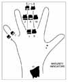
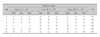
Skeletal Maturity Indicators (SMIs) were assigned by selecting six anatomical sites from the first, third and fifth phalanges based on the Skeletal Maturation Assessment (SMA) developed by Fishman9 (Fig 1).
Only the samples with an SMI Level of 6 or higher were included in this study, because the number of samples with SMI Levels of 5 or lower was minor.
Correlation between Demirjian Index and age at menarche
Correlations between the Demirjian Index and the age at menarche were examined in 224 out of the 270 female subjects whose ages at menarche were confirmed. The first radiograph was taken within six months after menarche, and the next radiographs were taken at six-month intervals.
Statistical analysis
(1) The median value, minimum value and maximum value of SMI, CVMI and chronologic age were calculated at each stage of the Demirjian Index.
(2) The Pearson correlation analysis was used to verify the relationship among the SMI, the CVMI and the Demirjian Index.
(3) The Spearman rank order correlation analysis was used to assess the relationship between age at menarche and third molar development.
(4) To evaluate the variations of third molar development among malocclusion classes, median, minimum, and maximum values of the Demirjian Index at every SMI stage for each malocclusion class were determined followed by a Kruskal-Wallis Test for the verification of variations.
RESULTS
Changes in chronological age along with the development of the third molar
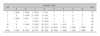
The median values were SMI Stage 10 and CVMI Stage 5 at Demirjian Stage D, and the mean chronologic age was 13.5 years. All the samples were at SMI Stage 11 and CVMI Stage 6 at Demirjian Stage G and H where root formation is almost completed (Table 3).
Distribution of SMI and CVMI by Demirjian Index
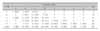
Deciding on the levels of SMI where a high proportion is occupied by Demirjian Stages from A to C is challenging because the number of samples in the stages is insignificant and has a diverse distribution. SMI Level 10 occupies nearly half (43.7%) of Demirjian Stage D. SMI Levels 10 and 11 are characterized by their concentration in Demirjian Stages beginning with E. Demirjian Stages G and H were all distinguished by SMI Level 11 (Table 4). In spite of the difference in ratio, Demirjian Stages from A to D displayed various distributions in CVMI Levels from 2 to 6. Demirjian Stages beginning with E were characterized by their concentration of CVMI Levels 5 and 6, which corresponded to the pattern of SMI distribution. Demirjian Stages G and H were all distinguished by CVMI Level 6 (Table 5).
Intercorrelations of SMI, CVMI and the Demirjian Index
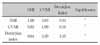
Upon examination of the intercorrelations between SMI and the Demirjian Index, and CVMI and the Demirjian Index, each showed a statistically significant correlation, with a slightly higher correlation existing between SMI and the Demirjian Index. The Spearman rank-order correlation coefficient between SMI and CVMI was 0.83, which was also statistically significant (p < 0.001) (Table 6).
Correlation between Demirjian Index and age at menarche
The Demirjian Index and age at menarche showed a weak (although statistically significant) correlation (r = 0.26). The period between the age at menarche and the age when the radiograph was taken was divided into six-month intervals (Table 7).
Correlation between the Demirjian Index and malocclusion type
Every sample was classified into malocclusion classes I, II, or III to compare the median, minimum and maximum value of the Demirjian Index for the SMI of each class. There was no statistically significant difference among the malocclusion classes (Table 8).
DISCUSSION
Many growth evaluation methods for precise prediction have been suggested.5-7 Dental maturity, in particular, has the advantage of easy evaluation during regular dental treatment. The relationship between dental maturity and bone maturity, however, shows diverse opinions among different studies.5-7,13-16 Third molars were excluded from most of the studies due to their assumed developmental variations. The aim of this study was to investigate the relationship between dental maturity and skeletal maturity using the development of the third molar in the mandible and also to evaluate the clinical value of the third molar.
Dental maturity can generally be determined by the stage of tooth eruption or the stage of tooth formation. The first disadvantage of this method is in determining its exact timing because it happens quickly. In addition, tooth eruption can be altered by local factors, systemic diseases and nutritional habits; the reliability of the method is questionable. Therefore, dental maturity in this study was determined by evaluating the stages of tooth formation, with an emphasis on Demirjian's method. The root of the third molar tends to be less divergent and more fused, making it harder to evaluate its development according to root length. The Demirjian Index evaluates the evident changes in the shape of the tooth without letting the tooth length affect the reliability of evaluation. Recent studies have verified that Demirjian's classification system shows the least intra-examiner and inter-examiner errors and a high correlation with biological age.19,20 Therefore, Demirjian's classification system was utilized in this study to assess third molar development.
Bolaños et al21 reported that the crown formation of the third molar was complete at the age of 14, and the root formation terminated at an average of 18.5 years based on their studies in a Spanish population. Moreover, Kullman et al22 indicated that the root formation began at about 15.1 years and was completed at an average age of 19.3 years. The crown formation was also completed by 13.5 years in this study, which resembled the ranges of previous studies.
The distribution of SMI was investigated at each Demirjian Stage. It is difficult to characterize the specific pattern of distribution due to its diversity in Demirjian Stages below D. All the samples were within SMI 6 to 8 at Demirjian Stage A. But this result was due to the insufficient sample size. If sample size becomes larger, the result will be more various just as at Stage B to D. On the contrary, the distribution of SMI was consistent at Demirjian Stage E to H even though sample size was small. Even if sample size becomes larger, this consistent pattern will be maintained. Only SMI Levels 10 and 11 were presented in Demirjian Stage E and F, and SMI Level 11 alone was seen in Demirjian Stage G and H. In the same way, the distribution of CVMI was investigated at each Demirjian stage, with similar patterns observed to those seen for SMI; manifold distribution up to Stage D, the presentation of only CVMI Levels 5 and 6 in Stages E and F, and the appearance of CVMI Level 6 in Stages G and H. Overall, the samples in Demirjian Stages E or above, the stage for the end of crown formation of the third molar and the onset of root formation, are predicted to occur at the higher levels of SMI Level 10 and CVMI Level 5.
There was a statistically significant correlation between the development of the third molar (Demirjian Index) and SMI (r = 0.64), and the Index and CVMI (r = 0.59), with a slightly higher correlation found between SMI and the Demirjian Index. SMI and CVMI also had a high correlation (r = 0.83) that corresponded with the results from Hassel and Farman's study.12 The findings also correspond to those of Demisch and Wartmann23 (r = 0.73) and Engström et al14 (r = 0.71), who reported a strong correlation between third molar formation and skeletal maturity. However, Lewis and Garn,15 Demirjian et al,6 and Krailassiri et al24 indicated that the relationship between the development of the third molar and bone maturity was poor. Moorrees et al25 indicated that excessively subdivided stages not only make it difficult to divide the development into precise stages, but also adversely affect the evaluation due to the insufficient amount of time to progress to the following stage. Thus, the division of tooth development into only a few stages is desirable for a meaningful comparison.
Different samples may also affect the result of correlations between third molars and bone maturity. Demisch and Wartmann23 stated that among white children, the correlation between third molars and bone maturity extended to 0.8, while Thai kids showed only a 0.3 correlation using the same method.24 Mincer et al26 studied 823 U.S. citizens between the ages of 14.1 and 24.9 years. No significant difference in the development of the third molar was observed with regard to race. However, Gorgani et al27 examined 229 black and 221 white U.S. citizens between 6 and 14 years of age, and determined that third-molar crown mineralization was completed about a year earlier in black Americans than in whites. The comparison of third-molar mineralization among Germans, Japanese and South Africans conducted by Olze et al19 using Demirjian's classification system showed that the development completed first in South Africans, then in Germans, and finally in the Japanese participants.
Nanda28 suggested that there was a correlation between age at menarche and dental maturity (r = 0.59). Demirjian et al6 maintained that dental development was independent and that dental maturity had a low correlation with skeletal and sexual maturity due to the predominant accuracy of the dental development index over others. Garn et al29 stated that although a correlation was present between dental development and skeletal and sexual maturity in general, the development of the third molar was independent, resulting in a weak correlation with skeletal and sexual maturity. In this study, 224 out of 270 females with a verified age at menarche showed little correlation between age at menarche and the development of third molars, which is in agreement with Garn et al's study29 (r = 0.26). Although it appeared to be higher than that seen in previous reports, the correlation coefficient is not enough to be meaningful for the correlation between age at menarche and the development of the third molar.
In this study, no statistically significant difference in the development of the third molar was present among Class I, II or III malocclusions. Nanda30 indicated that subjects with a skeletal open bite presented with an earlier onset of the adolescent growth spurt in the maturation of the facial bones than did those with a deep bite, and so did the dental maturation. Janson et al31 investigated the influence of facial type on dental development in subjects of the same chronological age. They showed that subjects with long faces tended to have an advanced dental maturation in comparison with short faces, which was expressed by a mean difference in dental age of six months. Our results showed no significant difference in third molar development among Class I, II or III malocclusions. The type of malocclusion should not affect the development of the third molar.
The revealed correlation between lower third molar development and skeletal maturity in this study will allow clinicians to use the mandibular third molar as an adjunctive tool to adolescent growth assessment in combination with cervical vertebrae and hand-wrist maturity evaluations. Individual variations should be taken into consideration when using the developmental stage of the third molar in growth evaluations because third molars are known for their many variations based on previous studies. This cross-sectional study has limitations on evaluating the results because the females in this study were in the pubertal growth period and mainly concentrated in Demirjian Stages C, D and E. Further longitudinal studies with a larger sample size are recommended for more accurate results.
CONCLUSION
The aims of this study were to evaluate a correlation between dental maturity and skeletal maturity of cervical vertebrae and hand-wrist and to estimate the clinical predictive value of the third molar. For this study, the samples were derived from panoramic, lateral cephalometric and hand-wrist radiographs of 270 female adolescents. Dental maturity (Demirjian Index) and skeletal maturity (skeletal maturation indicators) and cervical vertebrae maturation indicators were estimated from these radiographs. The results were as follows:
There was a significant correlation (r = 0.64) between the SMI and the Demirjian Index, and a similar correlation (r = 0.59) was observed between the CVMI and the Demirjian Index (p < 0.001).
If the Demirjian Index was above Stage E, SMI was above Stage 10 and CVMI was above Stage 5.
There was a weak correlation (r = 0.26) between menarche and the Demirjian Index (p < 0.001).
There was no significant difference in the Demirjian Index among Class I, II or III malocclusions.
Based on the results of this study, a dental maturity evaluation using the mandibular third molar would be an adjunctive tool to adolescent growth assessment in combination with cervical vertebrae and hand-wrist maturity evaluations. In particular, since it has been proven that a Demirjian Index score above Stage E means that the SMI Stage is above 10 and the CVMI Stage is above 5, judging the completion of growth may be possible when the beginning of root formation of the third molar is seen on a radiograph. Individual variations, however, should be taken into consideration to use the developmental stage of the third molar in growth evaluations because third molars are known for their many variations from previous studies.
 XML Download