

 PDF
PDF ePub
ePub Citation
 Print
Print


Abstract
Purpose
The first objective of this study was to compare the upper midface morpholgy, focusing on the soft tissues, between skeletal Class III maloccusion patients with midfacial depression and the norm. The second objective was to estimate and analyze the change in the upper midface soft tissues following surgical correction with maxillary advancement by Lefort I osteotomy and mandibular setback by bilateral sagittal split osteotomy (BSSRO).
Methods
The samples consisted of 34 adult patients (15 males and 12 females) with an average age of 21years, who had severe anteroposterior discrepancy with midfacial depression. These patients had received presurgical orthodontic treatment and surgical treatment which consisted of simultaneous Lefort I osteotomy and BSSRO.
Results
The correlation coefficient between changes in maxillary advancement and changes in Or' (soft tissue orbitale) was 0.599 (p < 0.05). Change in maxillary plane angle and vertical change of the maxilla were not correlated with the change in Or' (p < 0.05). The ratio of soft tissue change in Or' to maxillary advancement was 43.57 %, and 81.54 % in Sn. Regression equations between maxillary movement and Or' were devised. The r2 value was 0.476.
Figures and Tables
Fig 1
Landmarks and linear (Left) and angular (Right) measurements. S (Sella); N (Nasion); Or (Orbitale); Or' (Soft tissue Orbitale), the intersection point of the soft tissue on Orbitale and the FH plane; A (Subspinale, point A); Sn (Subnasale); G (Glabella); Nd, the intersection point of the FH plane and the dorsum of nose; S', the perpendicularly projected point from S onto the FH plane; N', the perpendicularly projected point from N onto the FH plane; A', the perpendicularly projected point from the point A onto the FH plane; Sn', the perpendicularly projected point from Sn onto the FH plane; G', the perpendicularly projected point from G onto the FH plane; S'-A'; S-N; S'-Or; S'-Or'; S'-G'; S'-Sn'; N'-Or; G'-Or'; A⊥FH, The distance from A point to FH plane; Or⊥NA, the distance from Or to NA line; Or'⊥GSn, the distance from Or' to GSn line; Sn⊥FH, the distance from Sn to FH plane.
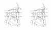
Table 1
Comparison of the mean values of soft tissue and skeletal tissue variables between skeletal Class III patients (T1) and normal occlusion in the previous report by Lee and Chung22 (male)
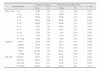
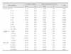
Table 2
Comparison of the mean values of soft tissue and skeletal tissue variables between skeletal Class III patients (T1) and normal occlusion in the previous report by Lee and Chung22 (female)

References
1. Vasir NS, Thompson RT, Davies TM. Dental and skeletal changes following sagittal split osteotomy for correction of mandibular prognathism. Eur J Orthod. 1991. 13:134–142.

2. McNeill RW, Proffit WR, White RP. Cephalometric prediction for orthodontic surgery. Angle Orthod. 1972. 42:154–164.
3. Burstone CJ, James RB, Legan H, Murphy GA, Norton LA. Cephalometrics for orthognathic surgery. J Oral Surg. 1978. 36:269–277.
4. Sung OJ, Kyung HM, Kwon OW, Sung JH. Cephalometric norms for orthognathic surgery. Korean J Orthod. 1989. 19:169–185.
5. Choe YK, Suhr CH. Hard and soft tissue changes after orthognathic surgery of mandibular prognathism. Korean J Orthod. 1993. 23:707–724.
6. Louis PJ, Austin RB, Waite PD, Mathews CS. Soft tissue changes of the upper lip associated with maxillary advancement in obstructive sleep apnea patients. J Oral Maxillofac Surg. 2001. 59:151–156.
7. Stella JP, Streater MR, Epker BN, Sinn DP. Predictability of upper lip soft tissue changes with maxillary advancement. J Oral Maxillofac Surg. 1989. 47:697–703.
8. Betts NJ, Vig P, Spalding P, Fonseca RJ. Changes in the nasal and soft tissues after surgical repositioning of the maxilla. Int J Adult Orthodon Orthognath Surg. 1993. 8:7–23.
9. Westermark AH, Bystedt H, von Konow L, Sällström KO. Nasolabial morphology after Le Fort I osteotomies. Effect of alar base suture. Int J Oral Maxillofac Surg. 1991. 20:25–30.
10. Mansour S, Burstone C, Legan H. An evaluation of soft-tissue changes resulting from Le Fort I maxillary surgery. Am J Orthod. 1983. 84:37–47.
11. Wolford LM. Discussion for lip-nasal aesthetics following Le Fort I osteotomy. Plast Reconstr Surg. 1988. 81:180–182.
12. Dann JJ 3rd, Fonseca RJ, Bell WH. Soft tissue changes associated with total maxillary advancement: a preliminary study. J Oral Surg. 1976. 34:19–23.
13. Choi YS, Son WS. A comparative study on the postsurgical changes between one jaw surgery and two-jaw surgery in skeletal Class III malocclusion patient. Korean J Orthod. 1997. 27:297–313.
14. Chang IH, Lee YJ, Park YG. A comparative study of soft tissue changes with mandibular one jaw surgery and double jaw surgery in Class III malocclusion. Korean J Orthod. 2006. 36:63–73.
15. Park HJ, Choi HR, Ryu SY. Soft tissue changes following bimaxillary surgery in skeletal Class III malocclusion patients. J Korean Assoc Maxillofac Plast Recontr Surg. 1998. 20:284–290.
16. Cho EJ, Yang WS. Soft tissue changes after double jaw surgery in skeletal Class III malocclusion. Korean J Orthod. 1996. 26:1–16.
17. Zide B, Grayson B, McCarthy JG. Cephalometric analysis for upper and lower midface surgery: Part II. Plast Reconstr Surg. 1981. 68:961–968.
18. Arnett GW, Bergman RT. Facial keys to orthodontic diagnosis and treatment planning. Part I. Am J Orthod Dentofacial Orthop. 1993. 103:299–312.
19. Arnett GW, Bergman RT. Facial keys to orthodontic diagnosis and treatment planning. Part II. Am J Orthod Dentofacial Orthop. 1993. 103:395–411.
20. Arnett GW, Jelic JS, Kim J, Cummings DR, Beress A, Worley CM Jr, Chung B, Bergman R. Soft tissue cephalometric analysis: diagnosis and treatment planning of dentofacial deformity. Am J Orthod Dentofacial Orthop. 1999. 116:239–253.
21. Kang SG, Lee YJ, Park YG. A comparative study of soft tissue profile between Korean and Caucasian young adults under NHP. Korean J Orthod. 2003. 33:323–337.
22. Lee EH, Chung IK. A lateral cephalometric analysis of midface focusing on zygomatic bone in Korean adults. J Korean Assoc Maxillofac Plast Recontr Surg. 1999. 21:353–359.
23. Riedel R. Esthetics and its relation to orthodontic therapy. Angle Orthod. 1950. 20:168–178.
24. Proffit WR, Phillips C, Turvey TA. Stability after surgical-orthodontic corrective of skeletal Class III malocclusion. 3. Combined maxillary and mandibular procedures. Int J Adult Orthodon Orthognath Surg. 1991. 6:211–225.
25. Leonard M, Walker GF. A cephalometric guide to the diagnosis of midface hypoplasia at the Le Fort II level. J Oral Surg. 1977. 35:21–24.
26. Boom LA. Perioral profile changes in orthodontic treatment. Am J Orthod. 1961. 47:371–379.
27. Denis KL, Speidel TM. Comparison of three methods of profile change prediction in the adult orthodontic patient. Am J Orthod Dentofacial Orthop. 1987. 92:396–402.
28. Gjørup H, Athanasiou AE. Soft-tissue and dentoskeletal profile changes associated with mandibular setback osteotomy. Am J Orthod Dentofacial Orthop. 1991. 100:312–323.
29. Betts NJ, Vig KW, Vig P, Spalding P, Fonseca RJ. Changes in the nasal and labial soft tissues after surgical repositioning of the maxilla. Int J Adult Orthodon Orthognath Surg. 1993. 8:7–23.
30. Willmot DR. Soft tissue profile changes following correction of class III malocclusions by mandibular surgery. Br J Orthod. 1981. 8:175–181.
31. Sarver DM, Ackerman MB. Dynamic smile visualization and quantification: Part 2. Smile analysis and treatment strategies. Am J Orthod Dentofacial Orthop. 2003. 124:116–127.
32. Han SY, Baik HS, Kim KD, Yu HS. Facial soft tissue measuring analysis of normal occlusion using three-dimensional CT imaging. Korean J Orthod. 2005. 35:409–419.
33. Bell WH. Modern practice in orthognathic and reconstructive surgery. 1992. St Louis: WB Saunders;2235–2297.
34. Abubaker AO, Sotereanos GC. Modified Le Fort I (maxillary-zygomatic) osteotomy: rationale, basis, and surgical technique. J Oral Maxillofac Surg. 1991. 49:1089–1097.
35. Nørholt SE, Sindet-Pedersen S, Jensen J. An extended Le Fort I osteotomy for correction of midface hypoplasia: a modified technique and results in 35 patients. J Oral Maxillofac Surg. 1996. 54:1297–1304.
36. Choung PH. A new midface plastic surgery technique: Development of intraoral Le Fort III, II and I composite osteotomy technique. J Korean Dent Assoc. 1997. 35:672–677.
37. Prendergast M, Schoenrock LD. Malar augmentation. Patient classification and placement. Arch Otolaryngol Head Neck Surg. 1989. 115:964–969.
38. Brusati R, Sesenna E, Raffaini M. On the feasibility of intraoral maxillo-malar osteotomy. J Craniomaxillofac Surg. 1989. 17:110–115.
39. Burstone CJ. The integumental profile. Am J Orthod. 1958. 44:1–25.
40. Wolford LM, Hilliard FW, Dugan DJ. Surgical Treatment Objectives: A systematic Approach to the Prediction Tracing. 1985. St Louis: Mosby;54–74.
41. Hohl TH, Epker BN. Macrogenia: A study of treatment results with surgical recommendations. Oral Surg Oral Med Oral Pathol. 1976. 41:545–567.
 XML Download