

 PDF
PDF ePub
ePub Citation
 Print
Print


Abstract
Objective
This study examined the craniofacial morphology of young patients in their prepubertal stage showing class I, II malocclusion, by analyzing lateral cephalograms, and analyzed its relationship with tongue position, tongue space, and airway space in order to ascertain the effects of nasopharyngeal airway and tongue morphology on the form of the malocclusion.
Methods
Seventy-six patients aging from 9 to 11 were divided into two groups depending on the ANB difference on the lateral cephalogram: Experimental group (Cl II malocclusion group) showing 0 ≤ ANB difference < 4.0; Control group (Cl I malocclusion group) showing 0 ≤ ANB difference < 4.0. The tongue space, space between palate and tongue, nasopharyngeal airway space and craniofacial morphology were compared between the two groups.
Results
Tongue space, palate-tongue space, nasopharyngeal airway space showed no significant differences between class I and class II malocclusion groups. Hyperdivergent faces were associated with smaller nasopharyngeal airway space. Longer anterior facial height and posterior facial height were associated with larger tongue space, and greater anterior facial height were associated with lower tongue position. Smaller nasopharyngeal airway space showed smaller tongue space.
Figures and Tables
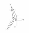
Fig 1
Landmarks and reference lines used in this study. Ar (Articulare), intersection between posterior cranial base surface and posterior border of condylar head and neck; Pt (Pterygoid point), the posterior point of the pterygopalatine fossa; ANS (anterior nasal spine), Anterior point of the maxilla; PNS (posterior nasal spine), Posterior point of the palatine bone; Me (Menton), the inferior point of the symphysis; Pm (Protuberance menti), the most superior point where the heavy cortical bone of the symphysis ends; Xi, midpoint of the ramus (Ricketts analysis); Rp, intersection between the posterior border of the ramus and the palatal plane; H1, intersection between posterior border of tongue and hyoid bone; H2, the most anterior point of the hyoid bone; T, the most anterior point of the outline of tongue; Palatal plane, a line passing through ANS and PNS.
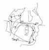
Fig 2
Spatial measurements used in this study. 1. Tongue space, area formed by superior and posterior border of tongue and T, Me, H2 and H1; 2. Palate-tongue space, space between tongue and palate from the line perpendicular to the palatal plane at the incisive foramen to the line perpendicular to the palatal plane at the PNS; 3. Nasopharyngeal airway space, area formed by Ar-Pt-PNS-Rp.
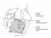
Fig 3
Anterior-posterior measurements used in this study. 4, A to N perpend, distance between A point and nasion perpendicular; 5, Facial convexity, angle formed by nasion, A point and pogonion; 6, Pog to N perpend, distance between pogonion and nasion perpendicular.
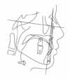
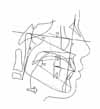
Fig 4
Vertical measurements used in this study. 7, anterior facial height (AFH), distance between nasion and menton; 8, posterior facial height (PFH), distance sella and gonion; 9, PFH/AFH; 10, cant of occlusal plane, an angle betweenthe FH plane and occlusal plane; 11, angle between facial plane and mandibular plane; 12, lower facial height, angle formed by ANS-Xi-Pm point.

References
1. Subtelny JD. Width of the nasopharynx and related anatomic structures in normal and unoperated cleft palate children. Am J Orthod. 1955. 41:889–909.

2. Tamari K, Shimizu K, Ichinose M, Nakata S, Tadahama Y. Relationship between tongue volume and lower dental arch sizes. Am J Orthod Dentofacial Orthop. 1991. 100:453–458.
3. Moyers RE. Etiology of Malocclusion. Handbook of Orthodontics. 1988. Chicago: Year Book Medical Publishers.
4. Solow B, Siersbaek-Nielsen S, Greve E. Airway adequacy, head posture, and craniofacial morphology. Am J Orthod. 1984. 86:214–223.
5. Linder-Aronson S. Adenoids. Their effect on mode of breathing and nasal airflow and their relationship to characteristics of the facial skeleton and the denition. A biometric, rhino-manometric and cephalometro-radiographic study on children with and without adenoids. Acta otolaryngol Suppl. 1970. 265:1–132.
6. Emslie RD, Massler M, Zwemer JD. Mouth breathing. 1. Etiology and effects; a review. J Am Dent Assoc. 1952. 44:506–521.
7. Subtelny JD, Sakuda M. Open-bite, Diagnosis and treatment. Am J Orthod. 1964. 50:337–358.
8. Melsen B, Stensgaard K, Pedersen J. Sucking habits and their influence on swallowing pattern and prevalence of malocclusion. Eur J Orthod. 1979. 1:271–280.
9. Lessa FC, Enoki C, Feres MF, Valera FC, Lima WT, Matsumoto MA. Breathing mode influence in craniofacial development. Rev Bras Otorrinolaringol. 2005. 71:156–160. (EnglEd).
10. Lopatiene K, Babarskas A. Malocclusion and upper airway obstruction. Medicina. 2002. 38:277–283. (Kaunas).
11. Houston WJ. The analysis of errors in orthodontic measurements. Am J Orthod. 1983. 83:382–390.
12. Proffit WR. Contemporary orthodontics. 2007. 4th ed. St Louis: Mosby;27–29.
13. Jaw TS, Sheu RS, Liu GC, Lin WC. Development of adenoids: a study by measurement with MR images. Kaohsiung J Med Sci. 1999. 15:12–18.
14. Valera FC, Travitzki LV, Mattar SE, Matsumoto MA, Elias AM, Anselmo-Lima WT. Muscular, functional and orthodontic changes in pre school children with enlarged adenoids and tonsils. Int J Pediatr Otorhinolaryngol. 2003. 67:761–770.
15. Niikuni N, Nakajima I, Akasaka M. The relationship between tongue-base position and craniofacial morphology in preschool children. J Clin Pediatr Dent. 2004. 28:131–134.
16. Cooper BC. Nasorespiratory function and orofacial development. Otolaryngol Clin North Am. 1989. 22:413–441.
17. Mergen DC, Jacobs RM. The size of Nasopharynx associated with normal occlusion and class II malocclusion. Angle Orthod. 1970. 40:342–346.
18. Behlfelt K, Linder-Aronson S, McWilliam J, Neander P, Laage-Hellman J. Cranio-facial morphology in children with and without enlarged tonsils. Eur J Orthod. 1990. 12:233–243.
19. Linder-Aronson S. Respiratory function in relation to facial morphology and the dentition. Br J Orthod. 1979. 6:59–71.
20. Hopkin GB. Neonatal and adult tongue dimensions. Angle Orthod. 1967. 37:132–133.
21. Lowe AA, Takada K, Yamagata Y, Sakuda M. Dentoskeletal and tongue soft-tissue correlates : A cephalometric analysis of rest position. Am J Orthod. 1985. 88:333–341.
22. Subtelny JD. The significance of adenoid tissue in orthodontia. Angle Orthod. 1954. 24:59–69.
23. Yoo E, Murakami S, Takada K, Fuchihata H, Sakuda M. Tongue volume in human female adults with mandibular prognathism. J Dent Res. 1996. 75:1957–1962.
24. Eifert DF. A roentogenographic cephalometric study of the tongue. Am J Orthod. 1960. 46:226–227.
 XML Download