

 PDF
PDF ePub
ePub Citation
 Print
Print


Abstract
Objective
This study analyzed the morphologic changes of the fourth cervical vertebra body to determine the skeletal age of orthodontic patients during growth.
Methods
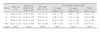
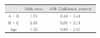
Eighty-one female patients aged from 11 to 14 who had cephalograms taken on the same day were examined. The subjects were divided into three groups depending on the depth of the concavity of the lower border of the fourth cervical vertebra (Group A: less than 1.05 mm, Group B: 1.05 - 2.07 mm, Group C: greater than 2.07 mm). Menarcheal timing, SMI stage, length, width and ratio of length and width of the fourth cervical vertebra body were analyzed and the following results were obtained.
Results
The average SMI stage of group A,B and C were 5.67 ± 2.57, 8.73 ± 2.41, and 10.00 ± 1.47, respectively. Length, width, ratio of length and width, and SMI stage were greater in group B than group A and in group C than group B. Mean menarcheal timing was 11.64 ± 0.92 years. Concavity depth, length, width, ratio of length and width showed a significant positive correlation with SMI stage, especially with the concavity depth.
Figures and Tables
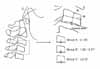
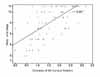
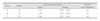
Table 1
Distribution of subjects by concavity depth of the inferior border at the 4th cervical vertebra


References
1. Bjork A, Helm S. Prediction of the age of maximum pubertal growth in body height. Angle orthod. 1967. 37:134–143.
2. Cha DS, Cha KS. A study on the comparisons between dental calcification and skeletal maturity. Korean J Orthod. 1994. 24:841–849.
3. Baume RM, Buschang PH, Weinstein S. Stature, head height and growth of the vertical face. Am J Orthod. 1983. 83:477–484.

4. Demirijian A, Buschang PH, Tanguay R, Patterson DK. Inter relationships among measures of somatic, skeletal, dental, and sexual maturity. Am J Orthod. 1985. 88:433–438.
5. Chapman SM. Ossification of the adductor sesamoid and the adolescent growth spurt. Angle Orthod. 1972. 42:236–244.
6. Liliequst B, Lundburg M. A methodological investigation. Acta Radio. 1971. 11:97–112.
7. Nanda SK. Prediction of facial growth using different biologic criteria in females. Craniofacial Growth Series, 20. 1986. Center for Human Growth and Development. Univ. of Michigan.
8. Lamparski DG. Skeletal age assessment utilizing cervical vertebrae. Master of dental thesis Pittsburgh Univ of Pittsburgh School of Dental Medicine. 1972.
9. Hassel B, Farman AG. Skeletal maturation evaluation using cervical vertebrae. Am J Orthod Dentofacial Orthop. 1995. 107:58–66.
10. Fishman LS. Radiographic evaluation of skeletal maturation; a clinically oriented method based on hand wrist films. Angle Orthod. 1982. 52:88–112.
11. Garcia-Fernandez P, Torre H, Flores L, Rea J. The cervical vertebrae as maturational indicators. J Clin Orthod. 1998. 32:221–225.
12. Krogman WM. The meaningful interpretation of growth and growth data by the clinician. Am J Orthod. 1958. 44:411–432.
13. Mito T, Sato K, Mitani H. Cervical vertebral bone age in girls. Am J Orthod Dentofacial Orthop. 2002. 122:380–385.
14. Bjork A. Sutural growth of the upper face studied by the implant method. Rep Congr Eur Orthod Soc. 1964. 40:49–65.
15. Gandini P, Mancini M, Andreani F. A comparison of hand-wrist bone and cervical vertebral analyses in measuring skeletal maturation. Angle Orthod. 2006. 76:984–989.
16. San Roman P, Palma JC, Oteo MD, Nevado E. Skeletal maturation determined by cervical vertebrae development. Eur J Orthod. 2002. 24:303–311.
17. Chang YH, Chung KR. The study on the relationship between the menarche and the bone maturity of malocclusion group. Korean J Orthod. 1995. 25:415–423.
 XML Download