

 PDF
PDF ePub
ePub Citation
 Print
Print


Abstract
Objective
This study was designed to analyze the primary and secondary stability characteristics of orthodontic mini-screws of tapered design when compared with the cylinder mini-screw.
Methods
A total of 48 mini-screws were placed into the buccal alveolar bone of the mandible in 6 male beagle dogs. Comparison was made between tapered and cylinder type mini-screws (Biomaterials Korea, Seoul, Korea). Maximum insertion torque (MIT) was measured using a torque sensor (Mark-10, MGT 50, USA) during installation, and maximum removal torque (MRT) was recorded after 3 and 12 weeks of loading.
Results
Taper mini-screws showed a higher MIT value of 22.3 Ncm compared with cylinder mini-screw showing 13.6 Ncm (p < 0.001). The MRT of the taper mini-screw showed a significantly higher value of 9.1 Ncm than those of cylinder mini-screw of 5.7 Ncm at 3-weeks after installation (p < 0.05). However, there was no difference in the MRT value between the taper and cylinder mini-screws at 12 weeks of loading.
Figures and Tables
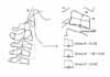
Fig 2
Schematic image for mini-screw insertion. a and b, Axial and sagittal images for the localization of mini-screws; c, force applied groups were reciprocally loaded by elastic-chain. FCS, force applied cylinder mini-screw; FTS, force applied taper mini-screw; CCS, control group of cylinder mini-screw; CTS, control group of taper mini-screw.

Fig 3
Timetable for placing mini-screw. w, weeks; FCS, force applied cylindrical mini-screw; FTS, force applied taper mini-screw; CCS, control group of cylindrical mini-screw; CTS, control group of taper mini-screw.
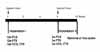
Fig 4
Insertion and removal torque (Ncm) for screw types. Statistically significant difference between periods by independent t-test; *p < 0.001.
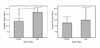
Fig 5
Graphs of insertion and removal torque for loading periods. Statistically significant difference between periods by independent t-test and Scheffe test, *p < 0.05; †p < 0.01; ‡p < 0.001.
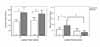
Fig 6
Graph of mobility change for loading periods. Cylinder-E, Cylinder type of experimental group; Cylinder-C, cylinder type of control group; Taper-E, taper type of experimental group; Taper-C, taper type of control group, *p < 0.05.
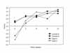
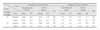
Table 2
Mobility change for loading periods
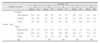
PT, Periotest value; SD, standard deviation; Sig, significance; NS, not significant; Statistically significant difference between cylinder and taper mini-screw by independent t-test, *p < 0.05. PT with -8 to +9 indicates clinically firm teeth or implant is well osseointegrated, PT over +10 indicates implant is not or not sufficiently osseointegrated (Shulte and Lukas24, 1992).
References
1. Chen CH, Chang CS, Hsieh CH, Tseng YC, Shen YS, Huang IY, et al. The use of microimplants in orthodontic anchorage. J Oral Maxillofac Surg. 2006. 64:1209–1213.

2. Buchter A, Wiechmann D, Koerdt S, Wiesmann HP, Piffko J, Meyer U. Load-related implant reaction of mini-implants used for orthodontic anchorage. Clin Oral Implants Res. 2005. 16:473–479.
3. Joos U, Buchter A, Wiesmann HP, Meyer U. Strain driven fast osseointegration of implants. Head Face Med. 2005. 1:6.
4. Huja SS, Litsky AS, Beck FM, Johnson KA, Larsen PE. Pull-out strength of monocortical screws placed in the maxilla and mandibles of dogs. Am J Orthod Dentofacial Orthop. 2005. 127:307–313.
5. Brown GA, McCarthy T, Bourgeault CA, Callahan DJ. Mechanical performance of standard and cannulated 4.0-mm cancellous bone screws. J Orthop Res. 2000. 18:307–312.
6. Heidemann W, Gerlach KL, Grobel KH, Kollner HG. Influence of different pilot hole sizes on torque measurements and pullout analysis of osteosynthesis screws. J Craniomaxillofac Surg. 1998. 26:50–55.
7. Miyawaki S, Koyama I, Inoue M, Mishima K, Sugahara T, Takano-Yamamoto T. Factors associated with the stability of titanium screws placed in the posterior region for orthodontic anchorage. Am J Orthod Dentofacial Orthop. 2003. 124:373–378.
8. Cheng SJ, Tseng IY, Lee JJ, Kok SH. A prospective study of the risk factors associated with failure of mini-implants used for orthodontic anchorage. Int J Oral Maxillofac Implants. 2004. 19:100–106.
9. Motoyoshi M, Hirabayashi M, Uemura M, Shimizu N. Recommended placement torque when tightening an orthodontic mini-implant. Clin Oral Implants Res. 2006. 17:109–114.
10. Meredith N. Assessment of implant stability as a prognostic determinant. Int J Prosthodont. 1998. 11:491–501.
11. Ueda M, Matsuki M, Jacobsson M, Tjellstrom A. Relationship between insertion torque and removal torque analyzed in fresh temporal bone. Int J Oral Maxillofac Implants. 1991. 6:442–447.
12. Frost HM. Bone's mechanostat: a 2003 update. Anat Rec A Discov Mol Cell Evol Biol. 2003. 275:1081–1101.
13. Frost HM. A brief review for orthopedic surgeons: fatigue damage (microdamage) in bone (its determinants and clinical implications). J Orthop Sci. 1998. 3:272–281.
14. Sowden D, Schmitz JP. AO self-drilling and self-tapping screws in rat calvarial bone: An ultrastructural study of the implant interface. J Oral Maxillofac Surg. 2002. 60:294–299.
15. Degidi M, Scarano A, Petrone G, Piattelli A. Histologic analysis of clinically retrieved immediately loaded titanium implants: a report of 11 cases. Clin Implant Dent Relat Res. 2003. 5:89–93.
16. Degidi M, Scarano A, Iezzi G, Piattelli A. Periimplant bone in immediately loaded titanium implants: histologic and histomorphometric evaluation in human. A report of two cases. Clin Implant Dent Relat Res. 2003. 5:170–175.
17. Melsen B, Lang NP. Biological reactions of alveolar bone to orthodontic loading of oral implants. Clin Oral Implants Res. 2001. 12:144–152.
18. Oh NH, Kim SH, Kook YA, Lee KH, Kang YG, Mo SS. Removal torque of sandblasted large grit and acid etched treated mini-implant. Korean J Orthod. 2006. 36:324–330.
19. Branemark R, Ohrnell L-O, Nilsson P, Thomsen P. Biomechanical characterization of osseointegration during healing: an experimental in vivo study in the rat. Biomaterials. 1997. 18:969–978.
20. Lee JH, Ryu HS, Lee DS, Hong KS, Chang BS, Lee CK. Biomechanical and histomorphometric study on the bone-screw interface of bioactive ceramic-coated titanium screws. Biomaterials. 2005. 26:3249–3257.
21. Park HS, Yen S, Jeoung SH. Histologic and biomechanical characteristics of orthodontic self-drilling and self-tapping microscrew implants. Korean J Orthod. 2006. 36:16–29.
22. Chen YJ, Chen YH, Lin LD, Yao CC. Removal torque of miniscrews used for orthodontic anchorage--a preliminary report. Int J Oral Maxillofac Implants. 2006. 21:283–289.
23. Simon H, Caputo AA. Removal torque of immediately loaded transitional endosseous implants in human subjects. Int J Oral Maxillofac Implants. 2002. 17:839–845.
24. Schulte W, Lukas D. The Periotest method. Int Dent J. 1992. 42:433–440.
 XML Download