

 PDF
PDF ePub
ePub Citation
 Print
Print


Abstract
Objective
The purpose of this study was to evaluate the mobility and ratio of the bone-implant contact (BIC) of a sandblasted, large grit and acid-etched (SLA) orthodontic micro-implant.
Methods
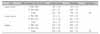
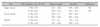
Ninety-six micro-implants (48 SLA and 48 machined) were implanted in the upper and lower buccal alveolar bone, and palatal bone of four beagle dogs. Two weeks after surgery, orthodontic force (150 - 200 g) was applied. Two beagles were sacrificed at 4-weeks and the other two at 12-weeks. Histomorphometric comparisons were made between the SLA experimental group and the machined micro-implant as a control group to determine the ratio of contact between the bone and implant. Micro-implant mobility was also evaluated using Periotest®.
Results
Periotest values showed no statistically significant difference in the upper alveolar and palatal bone between groups except for the lower buccal area. BIC in the upper buccal area showed no significant difference between groups both at 4-weeks and 12-weeks. However, both the groups showed a significant difference in BIC ratio in the rest of the experimental areas between 4 weeks and 12 weeks. The experimental group showed active bone remodeling around the bone-implant interface compared to the control group.
Figures and Tables
Fig 1
C-Implant components. A, C-Implant; B, surface-treated screw part; C, machined-surface screw part.
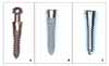
Fig 2
Application of force (150 - 200 g) by NiTi coil spring after 2 weeks of surgery in the buccal area (left), and in the palatal area (right).
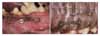
Fig 3
Photomicrograph of the machined surface micro-implant in the 4-week group (A) and 12-week group (B).

Fig 4
Photomicrograph of the surface treated micro-implant in the 4-week group (A, C) and 12-week group (B, D).
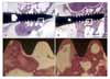
Fig 5
A, Fluorescence microscope image of the surface treated micro-implant in the 4-week group. a, First osteon; b, second osteon. B, Fluorescence microscope image of the machined micro-implant surface screw in the 4-week group.
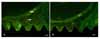
Fig 6.
A, Fluorescence microscope image of the surface treated micro-implant in the 12-week group. a, First osteon; b, second osteon; B, Fluorescence microscope image of the machined micro-implant in the 12-week group.

References
1. Roberts WE, Marshall KJ, Mozsary PG. Rigid endosseous implant utilized as anchorage to protract molars and close an atrophic extraction site. Angle Orthod. 1990. 60:135–152.
2. Im DH, Kim YS, Cho MA, Kim KS, Yang SE. Interdisplinary treatment of Class III malocclusion using mini-implant: problem-oriented orthodontic treatment. Korean J Orthod. 2007. 37:305–314.
3. Park HS. The skeletal cortical anchorage using titanium microscrew implants. Korean J Orthod. 1999. 29:699–706.
4. Park HS. A new protocal of the sliding mechanics with Micro-Implant Anchorage (M.I.A). Korean J Orthod. 2000. 30:677–685.
5. Cordioli G, Majzoub Z, Piatelli A, Scarano A. Removal torque and histomorphometric investigation of 4 different titanium surfaces: an experimental study in the rabbit tibia. Int J Oral Maxillofac Implants. 2000. 15:668–674.
6. Lim YJ, Oshida Y, Andres CJ, Barco MT. Surface characterizations of variously treated titanium materials. Int J Oral Maxillofac Implants. 2001. 16:333–342.
7. Cho SA, Park KT. The removal torque of titanium screw inserted in rabbit tibiatreated by dual acid etching. Biomaterials. 2003. 24:3611–3617.

8. Abrahamsson I, Berglundh T, Linder E, Lang NP, Lindhe J. Early bone formation adjacent to rough and turned endosseous implant surfaces. An experimental study in the dog. Clin Oral Implants Res. 2004. 15:381–392.
9. Lim SA, Cha JY, Hwang CJ. Comparison of insertion torque regarding changes in shape, diameter, and length of orthodontic miniscrews. Korean J Orthod. 2007. 37:89–97.
10. Song YY, Cha JY, Hwang CJ. Evaluation of insertion torque and Pull-out strength of mini-screws according to different thickness of artificial cortical bone. Korean J Orthod. 2007. 37:5–15.
11. Umemori M, Sugawara J, Mitani H, Nagasaka H, Kawamura H. Skeletal anchorage system for open-bite correction. Am J Orthod Dentofacial Orthop. 1999. 115:166–174.
12. Kyung SH, Lim JK, Park YC. The use of miniscrew as an anchorage for the orthodontic tooth movement. Korean J Orthod. 2001. 31:415–424.
13. Cho JH. Effects on orthodontic miniscrew implants according to the timing of force application. 2003. Seoul: Yonsei University;[thesis].
14. Sun SB, Kang YG, Kim SH, Mo SS, Kook YA. Influence of immediate loading on the removal torque value of mini-screws. Korean J Orthod. 2007. 37:400–406.
15. Chung KR, Kim SH, Kook YA. The C-orthodontic micro-implant. J Clin Orthod. 2004. 38:478–486.
16. Oh NH, Kim SH, Kook YA, Lee KH, Kang YG, Mo SS. Removal torque of sandblasted large grit, acid etched treated mini-implant. Korean J Orthod. 2006. 36:324–330.
17. Maino BG, Bednar J, Pagin P, Mura P. The spider screw for skeletal anchorage. J Clin Orthod. 2003. 37:90–97.
18. Yoon BS, Choi BH, Lee YU, Kim KN, Shim HB, Park JH. A study on Titaniuim Miniscrew as Orthodontic Anchorage; An experimental investigation in dogs. Korean J Orthod. 2001. 31:517–523.
19. Lim JW, Kim WS, Kim IK, Son CY, Byun HI. Three dimensional finite element method for stress distribution on the length and diameter of orthodontic miniscrew and cortical bone thickness. Korean J Orthod. 2003. 33:11–20.
20. Huja SS, Litsky AS, Beck FM, Johnson KA, Larsen PE. Pull-out strength of monocortical screws placed in the maxillae and mandibles of dogs. Am J Orthod Dentofacial Orthop. 2005. 127:307–313.
21. Kim SH, Cho JH, Chung KR, Kook YA, Nelson G. Evaluation of the removal torque values of surface-treated mini-implants after loading. Am J Orthod Dentofacial Orthop. 2008. 134:36–43.
22. Kim JW, Ahn SJ, Chang YI. Histomorphometric and mechanical analyses of the drill-free screw as orthodontic anchorage. Am J Orthod Dentofacial Orthop. 2005. 128:190–194.
23. Gurgel BC, Goncalves PF, Pimentel SP, Nociti FH, Sallum EA, Sallum AW, et al. An oxidized implant surface may improve bone-to-implant contact in pristine bone and bone defects treated with guided bone regeneration: an experimental study in dogs. J Periodontol. 2008. 79:1225–1231.
24. Iamoni F, Rasperini G, Trisi P, Simion M. Histomorphometric analysis of a half hydroxyapatite-coated implant in humans: a pilot study. Int J Oral Maxillofac Implants. 1999. 14:729–735.
25. Sennerby L, Thomsen P, Ericson LE. A morphometric and biomechanic comparison of titanium implants inserted in rabbit cortical and cancellous bone. Int J Oral Maxillofac Implants. 1992. 7:62–71.
26. Costa A, Raffaini M, Melsen B. Miniscrews as orthodontic anchorage: a preliminary report. Int J Adult Orthodon Orthognath Surg. 1998. 13:201–209.
27. Melsen B, Verna C. A rational approach to orthodontic anchorage. Prog Orthod. 1999. 1:10–22.
 XML Download