

 PDF
PDF ePub
ePub Citation
 Print
Print


Abstract
Objective
The aim of this study was to evaluate the remineralization of interdentally stripped teeth after fluoride gel or hydroxyapatite paste application.
Methods
After interdental stripping, 1.23% fluoride gel or 10% hydroxyapatite paste was applied three times a day, with a duration of four minutes, for a week. Scanning electron microscopy (SEM) and energy dispersive X-ray spectroscopy (EDS) were used to compare the change of surface contents and crystal structures before and after the application of fluoride gel or hydroxyapatite paste.
Results
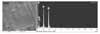
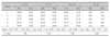
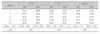
EDS analysis indicated that calcium contents were increased after 10% HAp paste application on stripped enamel (p < 0.01). SEM view showed that enamel surfaces in groups of 1.23% APF gel or 10% HAp paste application were smoother than those of control group, which was regarded as filling of the pore structure. And pores between crystal structures in groups treated with 1.23% APF gel or 10% HAp paste were smaller than those of control group.
Figures and Tables
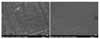 | Fig 1Surface appearance change of stripped enamel 7 days after acidulated phosphate fluoride (APF) gel application (SEM micrograph, × 250). A, Stripped enamel; B, 7 days after 1.23% APF gel application.
|
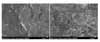 | Fig 2Crystal structure change of stripped enamel 7 days after acidulated phosphate fluoride (APF) gel application (SEM micrograph, × 50 k). A, Stripped enamel; B, 7 days after 1.23% APF gel application.
|
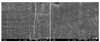 | Fig 3Surface appearance change of stripped enamel 7 days after 10% hydroxy apatite (HAp) paste application (SEM micrograph, × 250). A, Stripped enamel; B, 7 days after 10% HAp paste application.
|
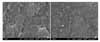 | Fig 4Crystal structure change of stripped enamel 7 days after 10% hydroxy apatite (HAp) paste application (SEM micrograph, × 50 k). A, Stripped enamel; B, 7 days after 10% HAp paste application.
|
References
1. Bolton WA. Disharmony in tooth size and its relation to the analysis and treatment of malocclusion. Angle Orthod. 1958. 28:113–130.
2. Tuverson DL. Anterior interocclusal relations. Part I. Am J Orthod. 1980. 78:361–370.

3. Sheridan JJ. Air-rotor stripping update. J Clin Orthod. 1987. 21:781–788.
4. Boese LR. Fiberotomy and reproximation without lower retention: nine years in retrospect. Angle Orthod. 1980. 50:88–97.
5. Blake M, Bibby K. Retention and stability: a review of the literature. Am J Orthod Dentofacial Orthop. 1998. 114:299–306.
6. Piacentini C, Sfondrini G. A scanning electron microscopy comparison of enamel polishing methods after air-rotor stripping. Am J Orthod Dentofacial Orthop. 1996. 109:57–63.
7. Radlanski RJ, Jäger A, Schwestka R, Bertzbach F. Plaque accumulations caused by interdental stripping. Am J Orthod Dentofacial Orthop. 1988. 94:416–420.
8. Twesme DA, Firestone AR, Heaven TJ, Feagin FF, Jacobson A. Air-rotor stripping and enamel demineralization in vitro. Am J Orthod Dentofacial Orthop. 1994. 105:142–152.
9. Arman A, Cehreli SB, Ozel E, Arhun N, Cetinşahin A, Soyman M. Qualitative and quantitative evaluation of enamel after various stripping methods. Am J Orthod Dentofacial Orthop. 2006. 130:131.
10. Joseph VP, Rossouw PE, Basson NJ. Orthodontic microabrasive reproximation. Am J Orthod Dentofacial Orthop. 1992. 102:351–359.
11. Kim KN. A study on the enamel surface texture and caries susceptibility in interdentally stripped teeth [thesis]. 2001. Gwangju: Chosun University.
12. Row J, Chun YS. A comparative study of roughness of enamel surface to various interdental enamel stripping methods in vitro. Korean J Orthod. 1999. 29:483–490.
13. Kim MY, Kwon HK, Kim BI. Remineralization effect of mouthrinse containing nano-hydroxyapatite by pH-cycling model. J Korean Acad Dent Health. 2007. 31:156–166.
14. Lee YE, Back HJ, Jeong SH, Choi YH, Song KB. Effect of hydroxyapatite containing dentifrice on the remineralization of early incipient carious lesion. J Korean Acad Dent Health. 2007. 31:305–318.
15. Barone M, Malpassi M. Clinical trial of a 15% supermicronized hydroxyapatite gel for dentin hypersensitivity. G Ital Endod. 1991. 5:43–47.
16. Huttemann RW, Strübel G, Rzekpa-Glinder V, Dönges H. Investigation of utility and mechanics of use of hydroxyapatite for therapy of hypersensitive tooth root. ZWR. 1989. 98:240–245.
17. Frazier MC, Southard TE, Doster PM. Prevention of enamel demineralization during orthodontic treatment: An in vitro study using pit and fissure sealants. Am J Orthod Dentofacial Orthop. 1996. 110:459–465.
18. Kim ME, Jung IL, Kim KY, Lee CY, Roh BD. In vivo quantitative analysis of remineralization effect of remineralization solution "R" of incipient enamel dental caries. J Korean Acad Conserv Dent. 2002. 27:175–182.
19. Kim SR, Hong SJ, Roh BD, Lee CY, Kum KY. The remineralizing effects of early enamel carious lesion by supersaturated buffer solution under pH cycling model. J Korean Acad Conserv Dent. 2001. 26:341–349.
20. Cha SW, Yoon TC, Park SH, Lee CY, Kum KY. Quantitative analysis of mineral change in the initial caries lesion using confocal laser scanning microscopy. J Korean Acad Conserv Dent. 2001. 26:1–8.
 XML Download