

 PDF
PDF ePub
ePub Citation
 Print
Print


Abstract
Objective
The purpose of this study was to compare the torque resistance to removal of sandblasted large grit and acid etched (SLA) surface treated orthodontic mini-implants and smooth surface orthodontic mini-implants as well as performing histologic observations.
Methods
Two groups of custom screw shaped orthodontic mini-implants (C-implant, 1.8 mm outer diameter × 9.5 mm length, Cimplant, Seoul, Korea) were designated. 22 SLA treated C-implants (SLA group) and 22 machined surface C-implants (machined group) were placed in the tibia metaphysis of 11 adult New Zealand white rabbits. Following a 6-week healing period, the rabbits were sacrificed. Subsequently, the C-implants were removed under reverse torque rotation with a digital torque measuring device and independent t-test was performed. Selected tissues were prepared for histologic observation.
Results
The
SLA group presented a higher mean removal torque value (6.286 Ncm) than the machined group (4.491 Ncm) which was statistically significant (p < 0.005). Histologic observation revealed a trend of more new bone formation in contact with the screw surface in the SLA group than the smooth group.
Figures and Tables
Fig. 1
Placement of C-implant. A, The machined C-implant placed on the right side of the tibia; B, the SLA treated C-implant placed on the left side of the tibia.
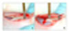
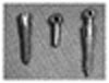
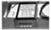
Fig. 6
Machined C-implant microphotograph. The C-implant was removed before histologic preparation. Fibrous encapsulation of bone marrow part of C-implant was observed (H-E staining, × 40).
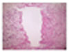
Fig. 7
Machined C-implant microphotograph. C-implant surface was covered with fibrous tissue. Note the new bone formation (arrow) within the fibrous tissue but not in contact with the implant surface (H-E staining, × 100).
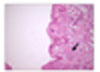
Fig. 8
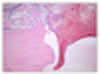
SLA C-implant microphotograph. C-implant was removed before histologic preparation. Deep part of the implant was covered by fibrous tissue but near cortical bone, new bone formation (dotted circle) was obvious (H-E staining, × 40).
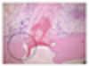
Fig. 9
SLA C-implant microphotograph. Higher magnification of Fig 8. Active new bone formation was observed with reversal line and cytoplasm abundant with osteoblasts (H-E staining, × 100).
References
1. Gainforth BL, Higley LB. A study of orthodontic anchorage possibilities in basal bone. Am J Orthod Oral Surg. 1945. 31:406–416.

2. Creekmore TD, Eklund MK. The possibility of skeletal anchorage. J Clin Orthod. 1983. 17:266–269.
3. Kanomi R. Mini-implant for orthodontic anchorage. J Clin Orthod. 1997. 31:763–767.
4. Costa A, Raffaini M, Melsen B. Miniscrew as orthodontic anchorage: a preliminary report. Int J Adult Orthodon Orthognath Surg. 1998. 13:201–209.
5. Melsen B, Verna C. A rational approach to orthodontic anchorage. Prog Orthod. 1999. 1:10–22.
6. Park HS. A new protocol of the sliding mechanics with micro-implant anchorage (M.I.A). Korean J Orthod. 2000. 30:677–685.
7. Park HS. Clinical study on success rate of micro screw implants for orthodontic anchorage. Korean J Orthod. 2003. 33:151–156.
8. Klokkevold PR, Nishimura RD, Adachi M, Caputo A. Osseointegration enhanced by chemical etching of the titanium surface. A torque removal study in the rabbit. Clin Oral Implants Res. 1997. 8:442–447.
9. Buser D, Nydegger T, Hirt HP, Cochran DL, Nolte LP. Removal torque values of titanium implants in the maxilla of miniature pigs. Int J Oral Maxillofac Implants. 1998. 13:611–619.
10. Cordioli G, Majzoub Z, Piatelli A, Scarano A. Removal torque and histomorphometric investigation of 4 different titanium surfaces: an experimental study in the rabbit tibia. Int J Oral Maxillofac Implants. 2000. 15:668–674.
11. Klokkevold PR, Johnson P, Dadgostari S, Caputo A, Davies JE, Nishimura RD. Early endosseous integration enhanced by dual acid etching of titanium: a torque removal study in the rabbit. Clin Oral Implants Res. 2001. 12:350–357.
12. Lee SJ, Chung KR. The effect of early loading on the direct bone-to-implant surface contact of the orthodontic osseointegrated titanium implant. Korean J Orthod. 2001. 31:173–185.
13. Cho SA, Jung SK. A removal torque of the laser-treated titanium implants in rabbit tibia. Biomaterials. 2003. 24:4859–4863.
14. Chung KR, Kim SH, Kook YA. The C-orthodontic micro-implant. J Clin Orthod. 2004. 38:478–486.
15. Chung K, Kim SH, Kook Y. C-orthodontic microimplant for distalization of mandibular dentition in Class III correction. Angle Orthod. 2005. 75:119–128.
16. Albrektsson T, Brånemark PI, Hansson HA, Lindström J. Osseointegration titanium implants. Requirements for ensuring a long-lasting, direct bone-to-bone implant anchorage in man. Acta Orthop Scand. 1981. 52:155–170.
17. Johansson C, Albrektsson T. Integration of screw implants in the rabbit: a 1-year follow-up of removal torque of titanium implants. Int J Oral Maxillofac Implants. 1987. 2:69–75.
18. Ivanoff CJ, Sennerby L, Johansson C, Rangert B, Lekholm U. Influence of implant diameters on the integration of screw implant. An experimental study in rabbits. Int J Oral Maxillofac Surg. 1997. 26:141–148.
19. Lim JW, Kim WS, Kim IK, Son CY, Byun HI. Three dimensional finite element method for stress distribution on the length and diameter of orthodontic miniscrew and cortical bone thickness. Korean J Orthod. 2003. 33:11–20.
20. Cha JY, Yoon TM, Hwang CJ. Insertion and removal torques according to orthodontic mini-screw design. Korean J Orthod. 2008. 38:5–12.
21. Carlsson I, Röstlund T, Albreksson B, Albrektsson T. Removal torques for polished and rough titanium implants. Int J Oral Maxillofac Implants. 1988. 3:21–24.
22. Darvell BW, Samman N, Luk WK, Clark RK, Tideman H. Contamination of titanium castings by aluminium oxide blasting. J Dent. 1995. 23:319–322.
23. Conchran DL, Nummikoski PV, Higginbottom FL, Hermann JS, Makins SR, Buser D. Evaluation of an endosseous titanium implant with sandblasted and acid-etched surface in the canine mandible: radiographic results. Clin Oral Implants Res. 1996. 7:240–252.
24. Baker D, London RM, O'Neal R. Rate of pull-out strength gain of dual-etched titanium implants: a comparative study in rabbits. Int J Oral Maxillofac Implants. 1999. 14:722–728.
25. Oh NH, Kim SH, Kook YA, Lee GH, Kang YG, Mo SS. Removal torque of sandblasted, large grit, acid etched treated mini-implant. Korean J Orthod. 2006. 36:324–330.
26. Buser D, Schenk RK, Steinemann S, Fiorellini JP, Fox CH, Stich H. Influence of surface characteristics on bone integration of titanium implants. A histomorphometric study in miniature pigs. J Biomed Mater Res. 1991. 25:889–902.
27. Costa A, Dalstra M, Melsen B. L'Aarhus anchorage system. Ortognatodonzia Italiana. 2000. 9:487–496.
 XML Download