

 PDF
PDF ePub
ePub Citation
 Print
Print


Abstract
Objective:
The purpose of this study was to evaluate the reproducibility of measurements representing asymmetry of the mandible and to identify which landmarks would be more useful in 3-dimensional (3D) CT imaging.
Methods:
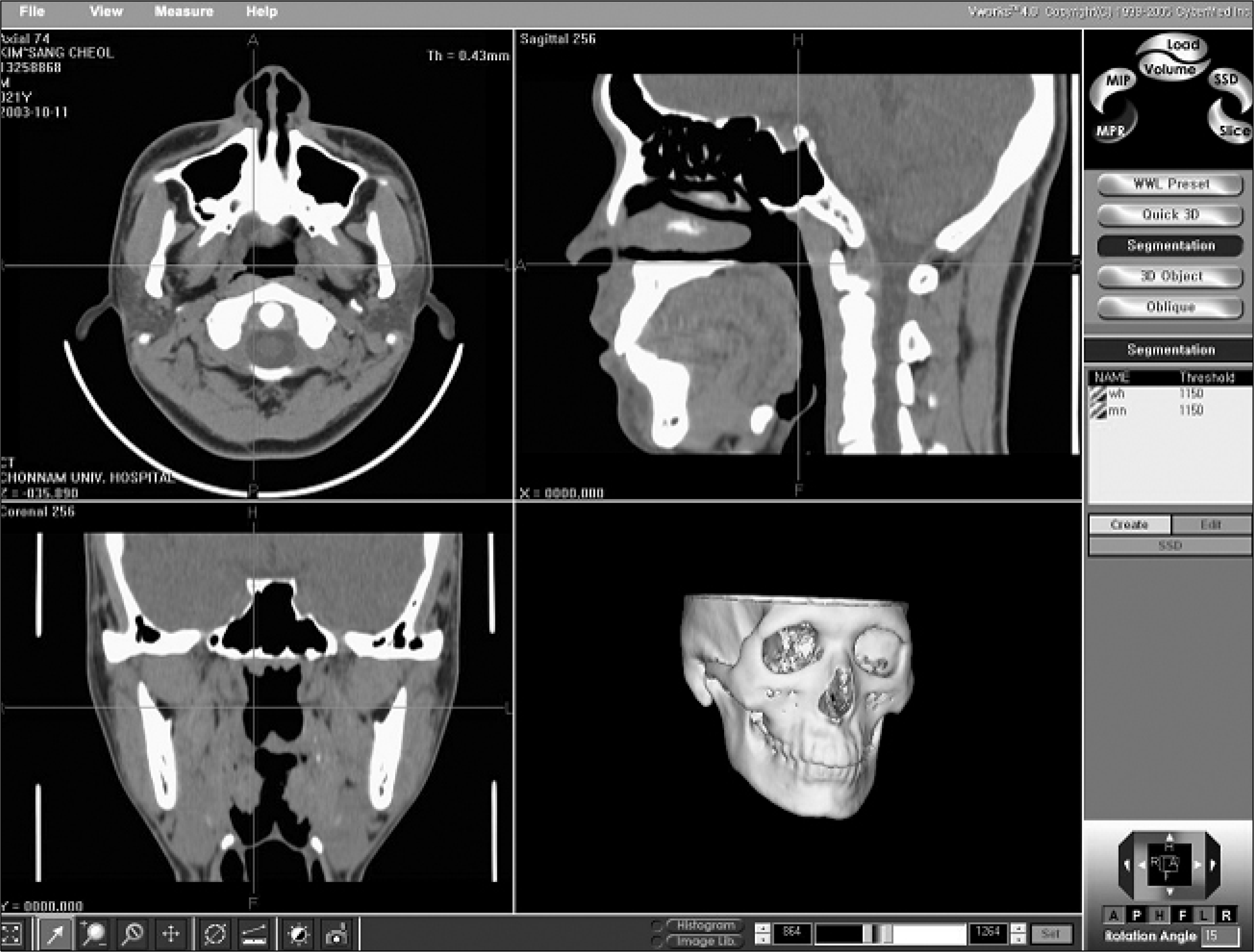
Facial CT images were obtained from forty normal occlusion individuals. Eighteen landmarks were established from the condyle, gonion, and menton areas, and 25 measurements were constructed to represent asymmetry of the mandible; 8 for ramus length, 12 for mandibular body length, 1 for condylar neck length, 2 for frontal ramal inclination, and 2 for lateral ramal inclination. Inter- and intra-examiner reproducibility of the measurements was evaluated.
Go to : 
REFERENCES
1.Ahn JS., Hwang HS. Relationship between perception of facial asymmetry and posteroanterior cephalometric measurements. Korean J Orthod. 2001. 31:489–98.
2.Shah SM., Joshi MR. An assessment of asymmetry in the normal craniofacial complex. Angle Orthod. 1978. 48:141–8.
3.Peck S., Peck L., Kataja M. Skeletal asymmetry in esthetically pleasing faces. Angle Orthod. 1991. 61:43–8.
4.Broadbent BH. A new x-ray technique and its application to orthodontia. Angle Orthod. 1931. 1:45–66.
5.Vogel CJ. Correction of frontal dimensions from head x-rays. Angle Orthod. 1967. 37:1–8.
6.Jarvinen S. A study of the factors causing differences in the relative variability of linear radiographic cephalometric measurements. Am J Orthod Dentofacial Orthop. 1987. 92:17–23.
7.Hatcher DC. Maxillofacial imaging. McNeill C, editor. editor.Science and Practice of Occlusion. Chicago: Quintessence Publishing;1997. p. 349–64.
8.Legrell PE., Nyquist H., Isberg A. Validity of identification of gonion and antegonion in frontal cephalograms. Angle Orthod. 2000. 70:157–64.
9.Berger H. Progress with basilar view cephalograms. Trans Eur Orthod Soc. 1964. 40:159–64.
10.Grayson B., Cutting C., Bookstein FL., Kim H., McCarthy JG. The three-dimensional cephalogram: theory, technique, and clinical application. Am J Orthod Dentofacial Orthop. 1988. 94:327–37.
11.Baumrind S., Moffitt FH., Curry S. Three-dimensional x-ray stereometry from paired coplanar images: a progress report. Am J Orthod. 1983. 84:292–312.

12.Baumrind S., Moffitt FH., Curry S. The geometry of three-dimensional measurement from paired coplanar x-ray images. Am J Orthod. 1983. 84:313–22.
13.Bookstein FL., Grayson B., Cutting CB., Kim HC., McCarthy JG. Landmarks in three dimensions: reconstruction from cephalograms versus direct observation. Am J Orthod Dentofacial Orthop. 1991. 100:133–40.
14.Kusnoto B., Evans CA., BeGole EA., de Rijk W. Assessment of 3-dimensional computer-generated cephalometric measurements. Am J Orthod Dentofacial Orthop. 1999. 116:390–9.
15.Koh EH., Lee KH., Hwang HS. Effects of vertical head rotation on the posteroanterior cephalometric measurements. Korean J Orthod. 2003. 33:73–84.
16.Vannier MW., Marsh JL., Warren JO. Three dimensional CT reconstruction images for craniofacial surgical planning and evaluation. Radiology. 1984. 150:179–84.
17.Dawood R. Digital radiology-a realistic prospect? Clin Radiol. 1990. 42:6–11.
18.Lill W., Solar P., Ulm C., Watzek G., Blahout R., Matejka M. Reproducibility of three-dimensional CT-assisted model production in the maxillofacial area. Br J Oral Maxillofac Surg. 1992. 30:233–6.
19.Altobelli DE., Kikinis R., Mulliken JB., Cline H., Lorensen W., Jolesz F. Computer-assisted three-dimensional planning in craniofacial surgery. Plast Reconstr Surg. 1993. 92:576–85.
20.Fuhrmann RA., Frohberg U., Diedrich PR. Treatment prediction with three-dimensional computer tomographic skull models. Am J Orthod Dentofacial Orthop. 1994. 106:156–60.
21.Darling CF., Byrd SE., Allen ED. Three-dimensional computed tomography imaging in the evaluation of craniofacial abnormalities. J Natl Med Assoc. 1994. 86:676–80.
22.Fuhrmann RA., Schnappauf A., Diedrich PR. Three-dimensional imaging of craniomaxillofacial structures with a standard personal computer. Dentomaxillofac Radiol. 1995. 24:260–3.
23.Fuhrmann R., Feifel H., Schnappauf A., Diedrich P. Integration of three-dimensional cephalometry and 3D-skull models in combined orthodontic/surgical treatment planning. J Orofac Orthop. 1996. 57:32–45.
24.Vannier MW., Hildebolt CF., Conover G., Knapp RH., Yokoyama-Crothers N., Wang G. Three-dimensional dental imaging by spiral CT. A progress report. Oral Surg Oral Med Pathol Oral Radiol Endod. 1997. 84:561–70.
25..Preda L., Di Maggio EM., Dore R., La Fianza A., Solcia M., Schifino MR, et al. Use of spiral computed tomography for multiplanar dental reconstruction. Dentomaxillofac Radiol. 1997. 26:327–31.
26.Cavalcanti MG., Vannier MW. Quantitative analysis of spiral computed tomography for craniofacial clinical applications. Dentomaxillofac Radiol. 1998. 27:344–50.
27.Quintero JC., Trosien A., Hatcher D., Kapila S. Craniofacial imaging in orthodontics: historical perspective, current status, and future developments. Angle Orthod. 1999. 69:491–506.
28.Chang HS., Baik HS. A proposal of landmarks for craniofacial analysis using three-dimensional CT imaging. Korean J Orthod. 2002. 32:313–25.
29.Hidelbolt CF., Vannier MW. Three-dimensional measurement accuracy of skull surface landmarks. Am J Phys Anthropol. 1988. 76:497–503.
30.Hidelbolt CF., Vannier MW., Knapp RH. Validation study of skull three-dimensional computerized tomography measurements. Am J Phys Anthropol. 1990. 82:283–94.
31.Williams FL., Richtsmeier JT. Comparison of mandibular landmarks from computed tomography and 3D digitizer data. Clin Anat. 2003. 16:494–500.
32.Kragskov J., Bosch C., Gyldensted C., Sindet-Pedersen S. Comparison of the reliability of craniofacial anatomic landmarks based on cephalometric radiographs and three-dimensional CT scans. Cleft Palate Craniofac J. 1997. 34:111–6.
33.Xia J., Wang D., Samman N., Yeung RW., Tideman H. Computer-assisted three-dimensional surgical planning and simulation: 3D color facial model generation. Int J Oral Maxillofac Surg. 2000. 29:2–10.
34.Edler R., Wertheim D., Greenhill D. Comparison of radiographic and photographic measurement of mandibular asymmetry. Am J Orthod Dentofacial Orthop. 2003. 123:167–74.
35.Graber TM. New horizons in case analysis-clinical cephalometrics. Am J Orthod. 1952. 38:603–24.
36.Ricketts RM. Provocations and perceptions in craniofacial orthopedics. Denver: Rocky Mountain, Inc.;1989. p. 797–803.
37.Sassouni V. Orthodontics in dental practice. St Louis: Mosby;1971. p. 330–7.
38.Gugino CF. An orthodontic philosophy. Denver: Rocky Mountain;1977. p. 1–2.
39.Peck H., Peck S. A concept of facial esthetics. Angle Orthod. 1970. 40:284–318.
Go to :
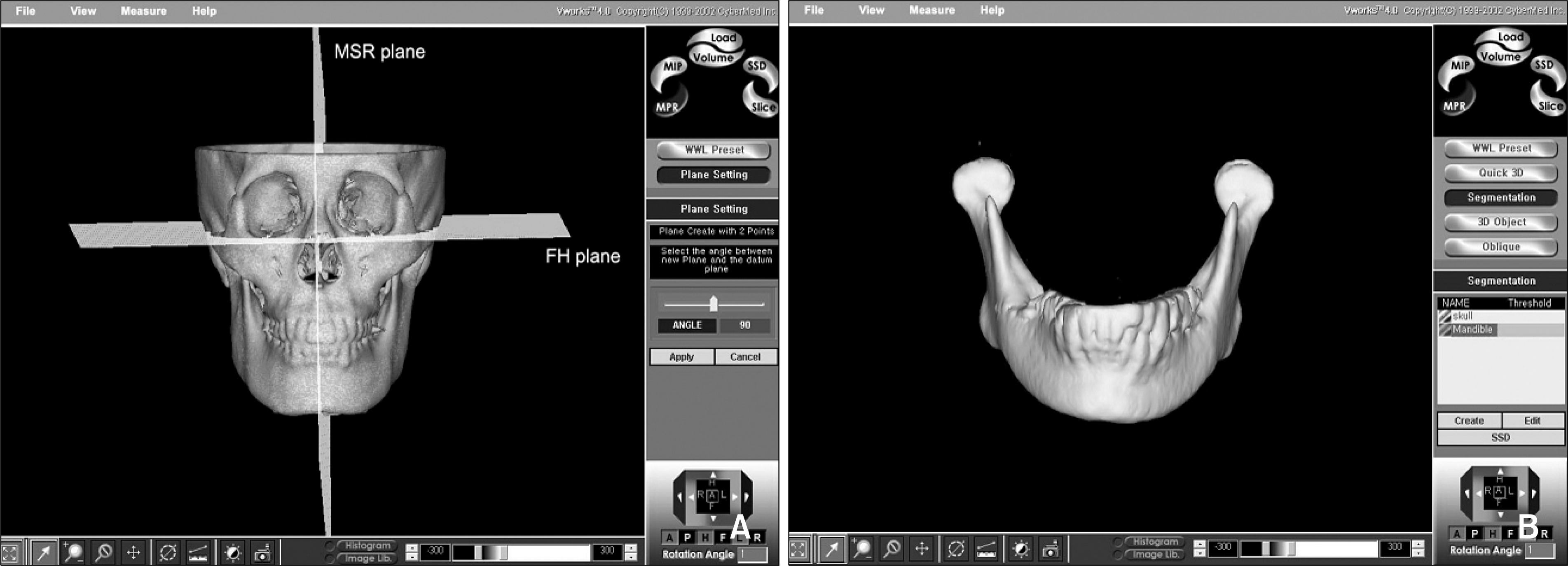 | Fig 2.
A, Construction of three-dimensional reference planes for measuring frontal and lateral ramal inclination (MSR plane; midsagittal plane, FH plane; Frankfort horizontal plane). B, The SOD file of the mandible was made in order to observe the mandible only. |
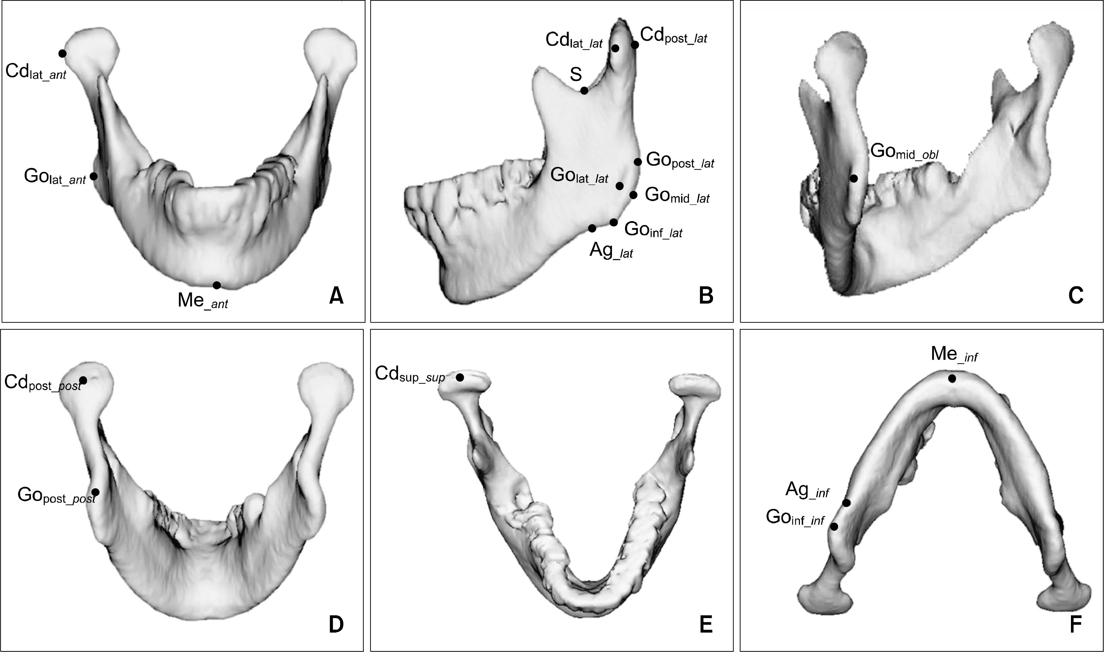 | Fig 3.Three-dimensional landmarks constructed. A, Anterior view; B, lateral view; C, postero-lateral view; D, posterior view; E, superior view; F, inferior view. The landmarks are described in Table 1. |
Table 1.
Description of three-dimensional landmarks constructed in this study
Table 2.
Linear and angular measurements constructed
Table 3.
Results of ANOVA test and intraclass correlation coefficient showing inter-examiner reproducibility of measurements (n = 40)
Table 4.
Post-hoc comparison by Tukey grouping in the measurements showing significant difference between exam-iners
Table 5.
Results of t-test showing intra-examiner reproducibility of measurements (n = 40)
Table 6.
Pearson correlation coefficient and reliability coefficient between first and second measurements showing intra-examiner reproducibility
 XML Download