

 PDF
PDF ePub
ePub Citation
 Print
Print


Abstract
Objective:
The purpose of this study was to investigate changes in the mandibular dental arch from presurgical orthodontic treatment and orthognathic surgery, and to evaluate the relationships between the pretreatment records and changes of mandibular dental arch in skeletal Class III malocclusion individuals.
Methods:
Lateral cephalometric radiographs and mandibular study models of 31 adults with skeletal class III malocclusion were taken and measured. All measurements were evaluated statistically by ANOVA, Scheffe's Post Hoc, and paired t-test, and correlation coefficients were evaluated.
Results:
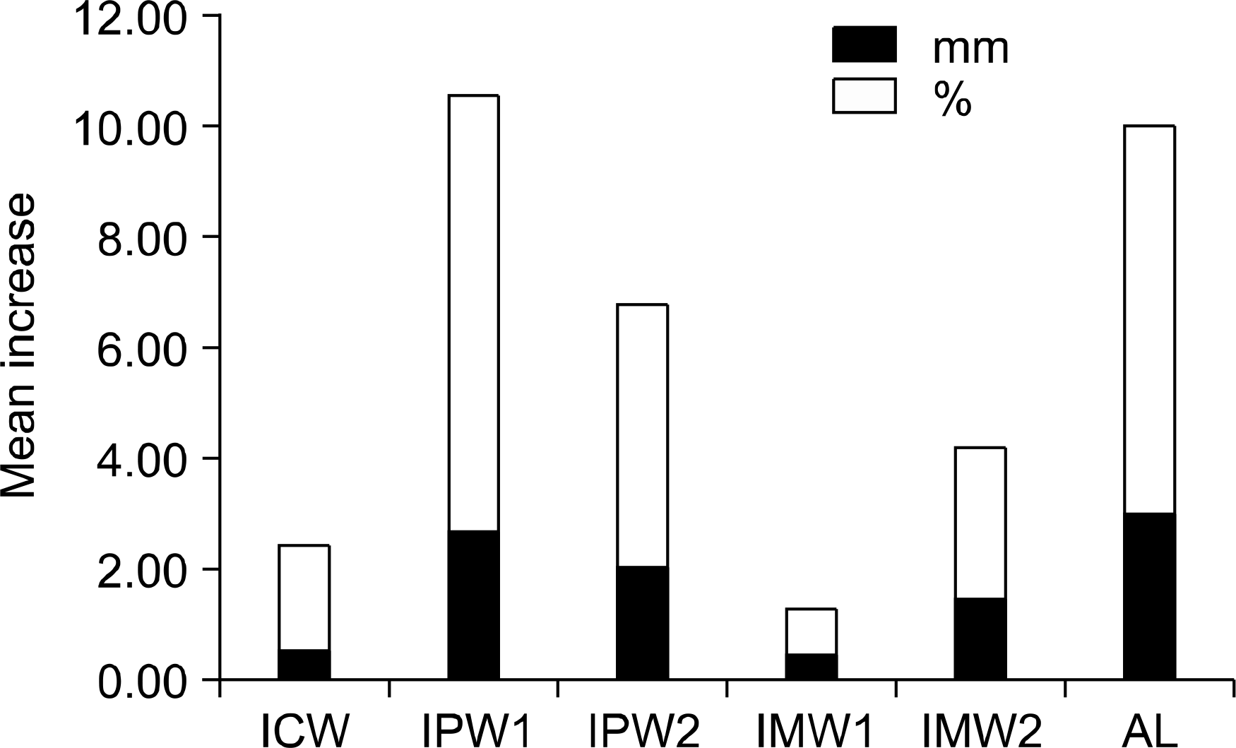
No significant difference in Mn-LMMC, Mn-LIE, Mn-MnOcc was detected between pretreatment and presurgical groups. Statistically significant but low correlations were demonstrated between the initial arch length discrepancy (ALD) and change in ICW, IPW1 (r = 0.492, 0.615) and change in arch length (r = 0.641). No association was seen between the initial depth of curve of Spee and change in mandibular incisor angle and arch width or arch length. Regression analysis showed that the amount of change for arch length and IPW1 could be explained by 64.0% and 75.8% of the pretreatment variables respectively.
Go to : 
REFERENCES
1.Yang SD. Surgical treatment objectives. J Korean Dent Assoc. 2007. 45:404–13.
2.Tae GC. Pre- and post-surgical orthodontic treatment. J Korean Dent Assoc. 2007. 45:413–22.
3.Montini RW., McGorray SP., Wheeler TT., Dolce C. Perceptions of orthognathic surgery patient's change in profile. A five-year follow-up. Angle Orthod. 2007. 77:5–11.
4.Worms FW., Isaacson RJ., Speidel TM. Surgical orthodontic treatment planning: profile analysis and mandibular surgery. Angle Orthod. 1976. 46:1–25.
5.Lee SJ., Hong SJ., Kim YH., Baek SH., Suhr CH. Effect of maxillary premolar extraction on transverse arch dimension in Class III surgical-orthodontic treatment. Korean J Orthod. 2005. 35:23–34.
6.Proffit WR., White RP., Sarver DM. Contemporary treatment of dentofacial deformity. St Louis: Mosby;2003. p. 245–67.
7.Wolford LM., Hilliard FW., Dugan DJ. Surgical treatment objective. St Louis: Mosby;1995. p. 11–74.
8.Steiner CC. Cephalometrics in clinical practice. Angle Orthod. 1959. 29:8–29.
9.Arnett GW., Jelic JS., Kim J., Cummings DR., Beress A., Worley CM Jr, et al. Soft tissue cephalometric analysis: diagnosis and treatment planning of dentofacial deformity. Am J Orthod Dentofac Orthop. 1999. 116:239–53.
10.Yang WS. Morphology of mandibular symphysis and positioning of lower incisors in the skeletal class III malocclusions. Korean J Orthod. 1985. 15:149–62.
11.Handelman CS. The anterior alveolus: its importance in limiting orthodontic treatment and its influence on the occurrence of iatrogenic sequelae. Angle Orthod. 1996. 66:95–109.
12.Wehrbein H., Bauer W., Diedrich P. Mandibular incisors, alveolar bone, and symphysis after orthodontic treatment. A retrospective study. Am J Orthod Dentofac Orthop. 1996. 110:239–46.
13.Hwang CJ., Kwon HJ. A study on the preorthodontic prediction values versus the actual postorthodontic values in class III surgery patients. Korean J Orthod. 2003. 33:1–9.
14.Kim SJ., Park SY., Woo HH., Park EJ., Kim YH., Lee SJ, et al. A study on the limit of orthodontic treatment. Korean J Orthod. 2004. 34:165–75.
15.Little RM. The irregularity index: a quantitative score of mandibular anterior alignment. Am J Orthod. 1975. 68:554–63.

16.Shannon KR., Nanda RS. Changes in the curve of Spee with treatment and at 2 years posttreatment. Am J Orthod Dentofacial Orthop. 2004. 125:589–96.
17.Bae GS., Son WS. Construction of an ideal set-up model for lingual orthodontic treatment. Korean J Orthod. 2005. 35:459–74.
18.Dahlberg G. Statistical methods for medical and biological students. New York: Interscience Publishers Inc.;1940. p. 122–32.
19.Bousaba S., Delatte M., Barbarin V., Faes J., De Clerck H. Pre-and post-surgical orthodontic objectives and orthodontic preparation. Rev Belge Med Dent. 2002. 57:37–48.
20.Swinnen K., Politis C., Willems G., De Bruyne I., Fieuws S., Heidbuchel K, et al. Skeletal and dento-alveolar stability after surgical-orthodontic treatment of anterior open bite: a retrospective study. Eur J Orthod. 2001. 23:547–57.
21.Lee HK., Son WS. A study on basal and dental arch width in skeletal Class III malocclusion. Korean J Orthod. 2002. 32:117–27.
22.Ishikawa H., Nakamura S., Iwasaki H., Kitazawa S., Tsukada H., Chu S. Dentoalveolar compensation in negative overjet cases. Angle Orthod. 2000. 70:145–8.
23.Jeon YJ., Park SB., Son WS. The correlation between dental compensation and craniofacial morphology in skeletal class III maloccusion. Korean J Orthod. 1997. 27:209–19.
24.Shim HY., Chang YI. Dentoalveolar compensation according to skeletal discrepancy in Normal occlusion. Korean J Orthod. 2004. 34:380–93.
25.Park SS., Kim HD., Lee DH., Jeon YM., Kim JG. Dentoalveolar characteristics according to facial types of class III malocclusion. Korean J Orthod. 2002. 32:33–42.
26.Jeong MH., Choi JH., Kim BH., Kim SG., Nahm DS. Soft tissue changes after double jaw rotation surgery in skeletal class III malocclusion. J Korean Assoc Oral Maxillofac Surg. 2006. 32:559–65.
27.Baek SH., Yang WS. A soft tissue analysis on facial esthetics of Korean young adults. Korean J Orthod. 1991. 21:131–70.
28.Robinson SW., Speidel TM., Isaacson RJ., Worms FW. Soft tissue profile change produced by reduction of mandibular prognathism. Angle Orthod. 1972. 42:227–35.
29.Capelozza Filho L., Martins A., Mazzotini R., da Silva Filho OG. Effects of dental decompensation on the surgical treatment of mandibular prognathism. Int J Adult Orthodon Orthognath Surg. 1996. 11:165–80.
30.Yang SD. Orthognathic surgery and orthodontic treatment goals. J Korean Found Gnatho-Orthod Res. 2003. 6:7–34.
31.Lee SJ., Kim TW., Nahm DS. Transverse implications of maxillary premolar extraction in class III presurgical orthodontic treatment. Am J Orthod Dentofacial Orthop. 2006. 129:740–8.
32.Willmot DR., Moss JP. Changes in the axial inclinations of upper and lower incisors after mandibular surgery in class III cases. J Maxillofac Surg. 1984. 12:163–6.
33.AlQabandi AK., Sadowsky C., BeGole EA. A comparison of the effects of rectangular and round arch wires in leveling the curve of Spee. Am J Orthod Dentofacial Orthop. 1999. 116:522–9.
34.Braun S., Hnat WP. Dynamic relationships of the mandibular anterior segment. Am J Orthod Dentofacial Orthop. 1997. 111:518–24.
35.Hemley S. Bite plates, their application and action. Am J Orthod Oral Surg. 1938. 24:721–36.
36.Strang RHM., Thompson WM. Case analysis. In: Textbook of Orthodontia. 4th ed.Lea and Febiger;Philadelphia: 1958. p. 335–61.
37.Chung TS., Sadowsky PL., Wallace DD., McCutcheon MJ. A three-dimensional analysis of mandibular arch changes following curve of Spee leveling in nonextraction orthodontic treatment. Int J Adult Orthodon Orthognath Surg. 1997. 12:109–21.
38.Im DH., Park HJ., Park JW., Kim JI., Chang YI. Surgical orthodontic treatment of skeletal class III malocclusion using mini-implant: correction of horizontal and vertical dental compensation. Korean J Orthod. 2006. 36:388–96.
39.Bailey LJ., Proffit WR., Blakey GH., Sarver DM. Surgical modification of long-face problems. Semin Orthod. 2002. 8:173–83.
Go to :
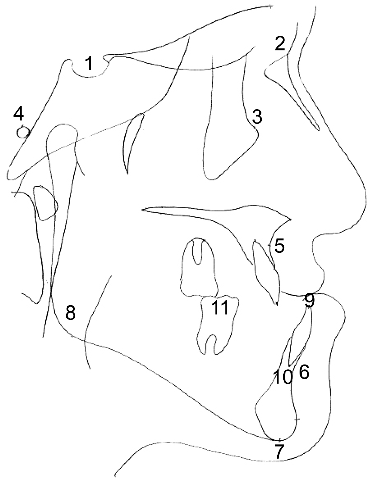 | Fig 1.Landmarks of lateral cephalometric films. 1, S (sella); 2, Na (nasion); 3, Or (orbitale); 4, Po (porion); 5, A (point A); 6, B (point B); 7, Me (menton); 8, Go (gonion); 9, LIE (lower incisor edge); 10, LIRA (lower incisor root apex); 11, LMMC (lower molar mesio-buccal cusp). |
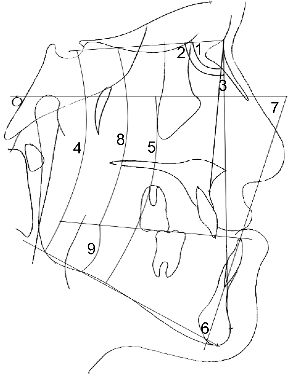 | Fig 2.Angular measurement of lateral cephalometric films. 1, SNA (SN plane to NA plane); 2, SNB (SN plane to NB plane); 3, ANB (NA plane to NB plane); 4, SN-Mn (SN plane to mandibular plane); 5, FH-Mn (FH plane to mandibular plane); 6, IMPA (lower incisor to mandibular plane); 7, FH-LI (FH plane to lower incisor); 8, SN-MnOcc (SN plane to mandibular occlusal plane); 9, Mn-MnOcc (mandibular plane to mandibular occlusal plane). |
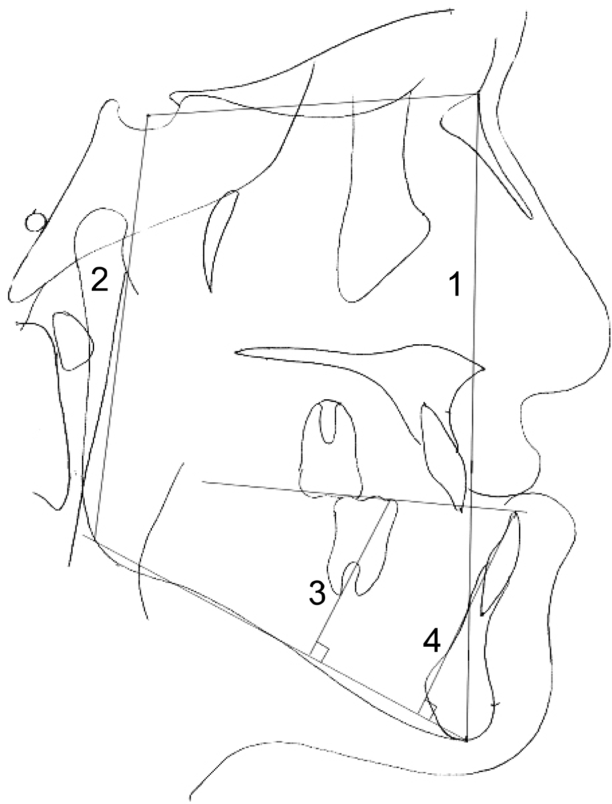 | Fig 3.Linear measurement of lateral cephalometric films. 1, AFH (anterior facial height); 2, PFH (posterior facial height); 3, Mn-LMMC (vertical distance between mandibular plane to lower molar mesio-buccal cusp); 4, Mn-LIE (vertical distance between mandibular plane to lower incisal edge). |
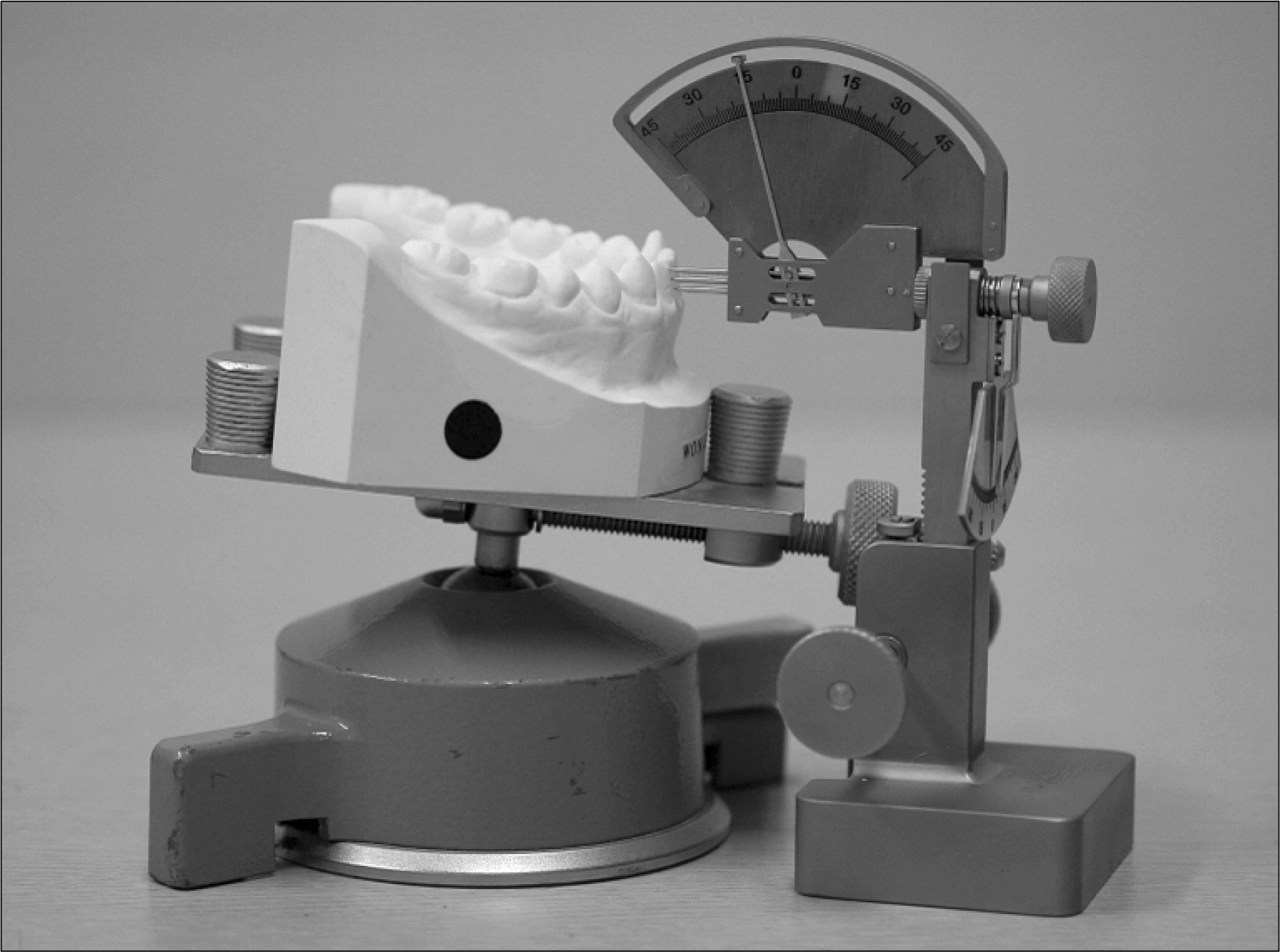
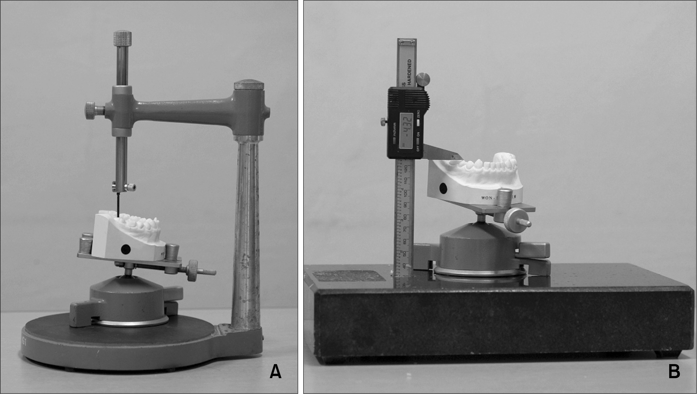 | Fig 4.Measurement of curve of Spee. A, Dental surveyor to allow measurement of angulation and inclination of each tooth and curve of Spee. B, surgical-orthodontic model calipers to measure curve of Spee. |
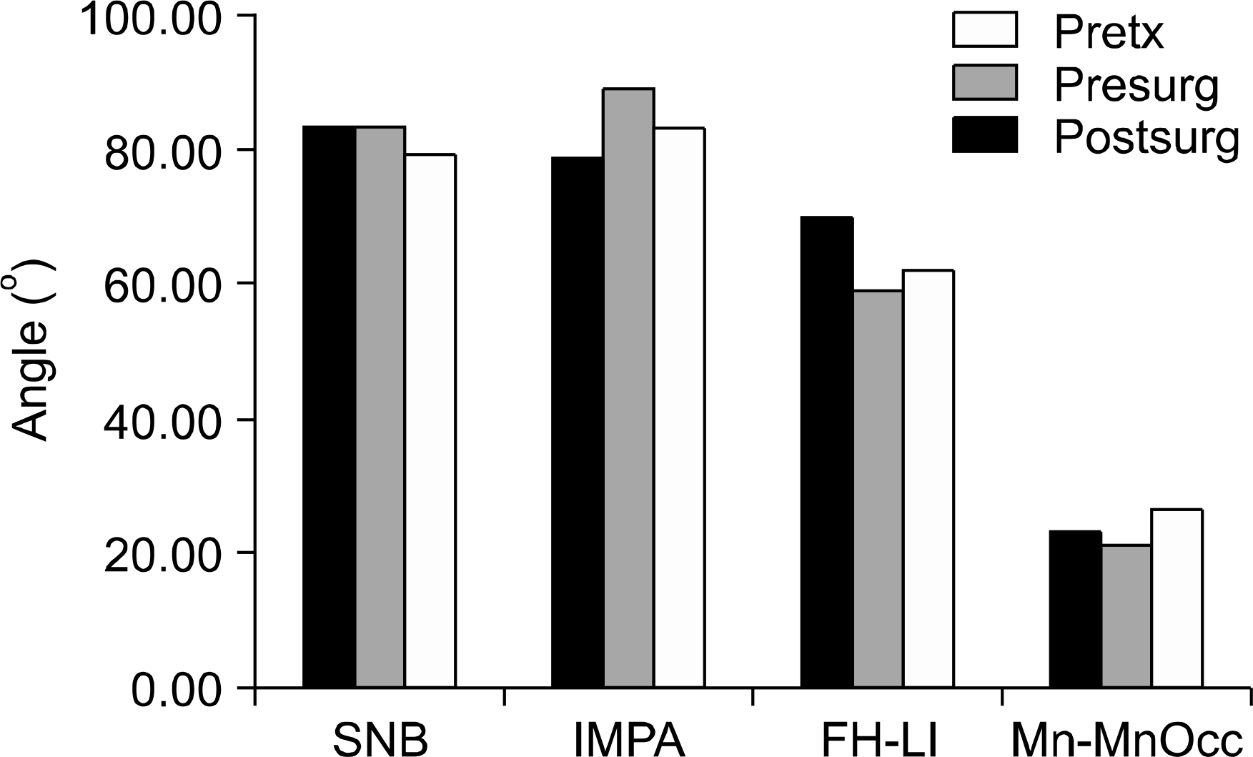 | Fig 6.Comparison of cephalometric measurements. Pretx, pretreatment; presurg, presurgical; postsurg, postsurgical. |
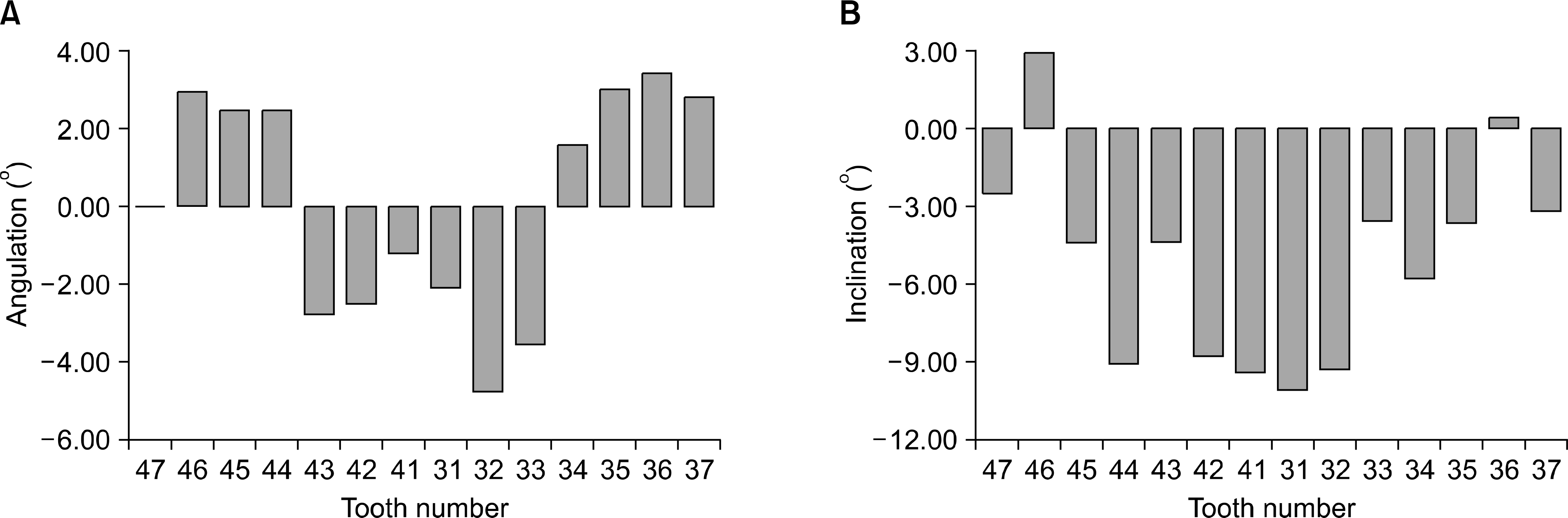 | Fig 8.(A) Difference in angulation between before and after treatment. Positive angulation values indicate distal tipping of clinical crown; negative values, mesial tipping. (B) Difference in inclination between before and after treatment. Positive inclination values indicate lingual tipping of clinical crown; negative values, labial tipping. |
Table 1.
Mean values and comparison of cephalometric measurements
| Measurement | Group | Mean ± | SD | Significance |
|---|---|---|---|---|
| Skeletal angular (o) | ||||
| SNA | T0 | 79.24a | 3.45 | |
| T1 | 79.40a | 3.14 | ∗ | |
| T2 | 80.43b | 4.15 | ||
| SNB | T0 | 83.58a | 3.75 | |
| T1 | 83.47a | 3.50 | † | |
| T2 | 79.30b | 3.66 | ||
| ANB | T0 | -3.35a | 3.50 | |
| T1 | -4.07a | 3.55 | † | |
| T2 | 1.13b | 3.44 | ||
| SN-Mn | T0 | 39.75 | 6.52 | |
| T1 | 39.77 | 6.15 | ||
| T2 | 42.52 | 5.82 | ||
| FH-Mn | T0 | 32.16 | 5.86 | |
| T1 | 31.85 | 5.72 | ||
| T2 | 34.54 | 5.00 | ||
| Skeletal linear (mm) | ||||
| AFH | T0 | 144.96 | 10.43 | |
| T1 | 145.33 | 9.71 | ||
| T2 | 142.37 | 9.25 | ||
| PFH | T0 | 90.01 | 7.46 | |
| T1 | 90.79 | 7.25 | ||
| T2 | 87.21 | 8.76 | ||
| PFH/AFH | T0 | 0.62 | 0.04 | |
| T1 | 0.63 | 0.04 | ||
| T2 | 0.61 | 0.05 | ||
| Dental angular (o) | ||||
| IMPA | T0 | 78.52a | 7.62 | |
| T1 | 89.03b | 8.31 | † | |
| T2 | 83.45c | 7.22 | ||
| FH-LI | T0 | 69.60a | 7.18 | |
| T1 | 59.18b | 8.00 | ∗ | |
| T2 | 62.26b | 8.05 | ||
| Dental linear (mm) | ||||
| Mn-LMMC | T0 | 38.09 | 3.52 | |
| T1 | 38.54 | 3.61 | ||
| T2 | 36.64 | 3.80 | ||
| Mn-LIE | T0 | 47.73 | 4.35 | |
| T1 | 48.90 | 4.14 | ||
| T2 | 49.00 | 3.86 | ||
| Occlusal angular(o) | ||||
| SN-MnOcc | T0 | 19.28 | 13.26 | |
| T1 | 18.25 | 5.12 | ||
| T2 | 15.94 | 4.85 | ||
| Mn-MnOcc | T0 | 22.91a | 4.62 | |
| T1 | 21.28a | 4.20 | † | |
| T2 | 26.31b | 4.73 | ||
Table 2.
Mean values and comparison between before treatment model and after treatment model in arch length discrepancy and curve of Spee of mandible (paired t-test)
Table 3.
Mean values and comparison between before treatment model and after treatment model in arch width and arch length of mandible (paired t-test)
Table 4.
Comparison between before treatment model and after treatment model in angulation and inclination of mandibular teeth (paired t-test)
Table 5.
Correlation coefficient analysis between pretreatment cephalometric measurements and each change (pretreatment to presurgical, n = 31)
| ΔPretx-Presurg | IMPA | FH-LI | ALD | II | COS | ICW- | IPW1 | IPW2 | IMW1 | IMW2 | AL |
|---|---|---|---|---|---|---|---|---|---|---|---|
| Pretx | |||||||||||
| SNB | 0.063 | 0.013 | 0.217 | -0.284 | 0.008 | -0.201 | 0.117 | -0.155 | -0.178 | -0.005 | 0.031 |
| SN-Mn | -0.033 | -0.048 | -0.209 | 0.203 | 0.083 | 0.083 | -0.212 | -0.024 | 0.090 | -0.036 | -0.054 |
| FH-Mn | -0.023 | -0.033 | -0.216 | 0.211 | 0.063 | 0.089 | -0.271 | -0.109 | 0.087 | -0.011 | -0.075 |
| AFH | 0.184 | -0.249 | -0.018 | 0.022 | 0.256 | 0.227 | -0.055 | 0.143 | 0.057 | -0.172 | 0.140 |
| PFH | 0.129 | -0.132 | 0.090 | -0.053 | 0.248 | 0.212 | 0.165 | 0.229 | 0.018 | -0.101 | 0.135 |
| PFH/AFH | -0.037 | 0.100 | 0.123 | -0.081 | 0.022 | 0.013 | 0.252 | 0.113 | -0.039 | 0.062 | 0.018 |
| IMPA | 0.236 | -0.199 | 0.060 | 0.065 | 0.107 | 0.145 | 0.101 | 0.346 | -0.070 | -0.185 | 0.328 |
| FH-LI | -0.275 | 0.271 | 0.072 | -0.183 | -0.173 | -0.211 | 0.079 | -0.307 | 0.027 | 0.217 | -0.280 |
| Mn-LMMC | 0.073 | -0.094 | 0.013 | 0.143 | 0.083 | 0.125 | -0.038 | 0.098 | 0.089 | 0.090 | 0.088 |
| Mn-LIE | 0.143 | -0.181 | 0.084 | 0.098 | 0.194 | 0.128 | -0.050 | 0.041 | -0.209 | -0.190 | 0.231 |
| SN-MnOcc | 0.258 | -0.297 | -0.219 | 0.035 | 0.460∗ | 0.050 | -0.156 | 0.333 | 0.042 | -0.173 | 0.007 |
| Mn-MnOcc | -0.181 | 0.152 | 0.154 | -0.156 | 0.250 | 0.047 | 0.105 | 0.016 | -0.299 | -0.255 | -0.041 |
Table 6.
Correlation coefficient analysis between pretreatment model measurements and each change (pretreatment to presurgical, n = 31)
| ΔPretx-Presurg | IMPA | FH-LI | ALD | II | COS | ICW | IPW1 | IPW2 | IMW1 | IMW2 | AL |
|---|---|---|---|---|---|---|---|---|---|---|---|
| Pretx | |||||||||||
| ALD | 0.351 | -0.329 | 0.996‡ | -0.635‡ | -0.198 | 0.492† | 0.615‡ | 0.287 | 0.287 | 0.197 | 0.641‡ |
| II | -0.312 | 0.283 | -0.765‡ | 0.958‡ | -0.045 | -0.425∗ | -0.509† | -0.347 | -0.438∗ | -0.088 | -0.595‡ |
| COS | 0.009 | -0.007 | 0.007 | -0.241 | 0.839‡ | 0.170 | -0.067 | 0.331 | -0.076 | -0.351 | 0.132 |
| Arch width | |||||||||||
| ICW | -0.016 | 0.005 | 0.387∗ | -0.171 | 0.156 | 0.707‡ | 0.212 | 0.365∗ | 0.015 | -0.036 | 0.537† |
| IPW1 | 0.241 | -0.206 | 0.587† | -0.413∗ | 0.015 | 0.425∗ | 0.775‡ | 0.507† | 0.170 | -0.028 | 0.540† |
| IPW2 | 0.242 | -0.217 | 0.273 | -0.309 | 0.450∗ | 0.471† | 0.437∗ | 0.870‡ | 0.265 | -0.234 | 0.429∗ |
| IMW1 | 0.124 | -0.103 | 0.202 | -0.310 | 0.271 | 0.376∗ | 0.294 | 0.490† | 0.571 | 0.204 | 0.247 |
| IMW2 | -0.102 | 0.118 | 0.158 | -0.214 | 0.230 | 0.261 | 0.170 | 0.276 | 0.442∗ | 0.346 | 0.070 |
| ICW-IMW1 | -0.144 | 0.111 | 0.154 | 0.160 | -0.133 | 0.275 | -0.104 | -0.164 | -0.576† | -0.245 | 0.249 |
| Arch length | 0.205 | -0.187 | 0.116 | 0.043 | 0.130 | 0.110 | -0.054 | 0.084 | -0.253 | -0.189 | 0.486† |
Table 7.
Correlation coefficient analysis between pretreatment model measurements (angulation) and each change (pretreatment to presurgical, n = 31)
| ΔPretx-Presurg | IMPA | FH-LI | ALD | II | COS | ICW | IPW1 | IPW2 | IMW1 | IMW2 | AL |
|---|---|---|---|---|---|---|---|---|---|---|---|
| Pretx | |||||||||||
| Angulation | |||||||||||
| 31 | 0.134 | -0.106 | 0.342 | -0.283 | -0.106 | 0.243 | 0.358∗ | 0.262 | 0.343 | 0.118 | 0.283 |
| 32 | 0.306 | -0.284 | 0.505† | -0.359∗ | 0.143 | 0.368∗ | 0.596‡ | 0.506† | 0.198 | -0.140 | 0.274 |
| 33 | -0.075 | 0.050 | -0.152 | 0.337 | -0.017 | -0.082 | -0.014 | -0.131 | -0.194 | -0.037 | -0.099 |
| 34 | -0.137 | 0.129 | -0.395∗ | 0.044 | 0.421∗ | -0.029 | -0.252 | 0.002 | -0.117 | -0.466† | -0.025 |
| 35 | -0.174 | 0.177 | -0.190 | 0.061 | 0.233 | 0.021 | -0.134 | 0.009 | 0.104 | -0.071 | -0.052 |
| 36 | -0.076 | 0.068 | 0.001 | -0.173 | 0.438∗ | 0.027 | 0.056 | 0.157 | 0.137 | -0.006 | 0.048 |
| 37 | 0.476† | -0.464† | 0.460† | -0.445∗ | 0.068 | 0.097 | 0.380∗ | 0.208 | 0.186 | -0.025 | 0.376∗ |
| 41 | 0.120 | -0.147 | 0.303 | -0.054 | -0.155 | 0.064 | 0.075 | 0.071 | 0.125 | 0.143 | -0.007 |
| 42 | 0.450∗ | -0.435∗ | 0.116 | -0.076 | 0.008 | -0.063 | 0.329 | 0.280 | 0.417∗ | 0.168 | 0.125 |
| 43 | -0.235 | 0.207 | -0.277 | 0.437∗ | -0.021 | -0.091 | -0.012 | -0.012 | -0.210 | -0.038 | -0.155 |
| 44 | -0.256 | 0.201 | -0.353 | 0.482† | 0.149 | 0.136 | -0.408∗ | -0.091 | -0.143 | 0.023 | -0.292 |
| 45 | -0.109 | 0.037 | -0.101 | 0.254 | 0.048 | 0.206 | -0.150 | -0.120 | 0.090 | 0.125 | -0.115 |
| 46 | 0.094 | -0.124 | 0.125 | -0.138 | 0.336 | 0.210 | -0.120 | -0.018 | 0.041 | 0.054 | 0.198 |
| 47 | 0.234 | -0.221 | 0.231 | -0.183 | 0.233 | -0.175 | -0.068 | -0.233 | -0.017 | 0.234 | 0.105 |
Table 8.
Correlation coefficient analysis between pretreatment model measurements (inclination) and each change (pretreatment to presurgical, n = 31)
| ΔPretx-Presurg | IMPA | FH-LI | ALD | II | COS | ICW | IPW1 | IPW2 | IMW1 | IMW2 | AL |
|---|---|---|---|---|---|---|---|---|---|---|---|
| Pretx | |||||||||||
| Inclination | |||||||||||
| 31 | 0.309 | -0.316 | 0.119 | -0.112 | 0.242 | 0.280 | 0.172 | 0.456∗ | -0.071 | -0.344 | 0.533† |
| 32 | 0.272 | -0.236 | 0.356∗ | -0.282 | 0.303 | 0.383∗ | 0.334 | 0.599‡ | 0.053 | -0.274 | 0.374∗ |
| 33 | -0.044 | 0.031 | -0.009 | 0.056 | 0.304 | 0.438∗ | 0.027 | 0.368∗ | -0.052 | -0.308 | 0.323 |
| 34 | 0.339 | -0.307 | 0.357∗ | -0.279 | 0.031 | 0.368∗ | 0.694‡ | 0.530† | 0.132 | -0.198 | 0.433∗ |
| 35 | 0.250 | -0.204 | 0.170 | -0.197 | 0.412∗ | 0.313 | 0.330 | 0.811‡ | 0.461† | -0.101 | 0.225 |
| 36 | 0.295 | -0.252 | 0.201 | -0.335 | 0.192 | 0.280 | 0.351 | 0.518† | 0.661‡ | 0.145 | 0.183 |
| 37 | 0.099 | -0.046 | 0.329 | -0.289 | 0.155 | 0.142 | 0.380∗ | 0.459† | 0.480† | 0.265 | 0.173 |
| 41 | 0.258 | -0.236 | 0.095 | -0.043 | 0.284 | 0.274 | 0.219 | 0.458† | -0.040 | -0.263 | 0.471† |
| 42 | 0.099 | -0.059 | 0.236 | -0.192 | 0.226 | 0.373∗ | 0.214 | 0.499† | 0.025 | -0.262 | 0.363∗ |
| 43 | 0.169 | -0.143 | 0.055 | -0.032 | 0.080 | 0.275 | 0.250 | 0.359∗ | 0.121 | -0.057 | 0.343 |
| 44 | 0.176 | -0.110 | 0.256 | -0.289 | 0.075 | 0.279 | 0.591‡ | 0.504† | 0.109 | -0.189 | 0.343 |
| 45 | 0.179 | -0.130 | 0.107 | -0.163 | 0.281 | 0.395∗ | 0.334 | 0.743‡ | 0.167 | -0.345 | 0.251 |
| 46 | -0.038 | 0.117 | 0.185 | -0.184 | -0.089 | 0.385∗ | 0.302 | 0.438∗ | 0.264 | 0.095 | 0.252 |
| 47 | 0.009 | 0.054 | 0.022 | -0.157 | 0.192 | 0.138 | 0.175 | 0.394∗ | 0.271 | 0.168 | -0.004 |
Table 9.
Correlation coefficient analysis between each change of measurements (pretreatment to presurgical, n = 31)
| IMPA | FH-LI | ALD | II | COS | ICW | IPW1 | IPW2 | IMW1 | IMW2 | AL | |
|---|---|---|---|---|---|---|---|---|---|---|---|
| IMPA | 1.000 | ||||||||||
| FH-LI | -0.978‡ | 1.000 | |||||||||
| ALD | 0.337 | -0.317 | 1.000 | ||||||||
| II | -0.259 | 0.230 | -0.664‡ | 1.000 | |||||||
| COS | -0.011 | 0.020 | -0.188 | -0.134 | 1.000 | ||||||
| ICW | 0.047 | -0.110 | 0.498† | -0.384∗ | 0.006 | 1.000 | |||||
| IPW1 | 0.273 | -0.256 | 0.630‡ | -0.492† | -0.206 | 0.484† | 1.000 | ||||
| IPW2 | 0.217 | -0.200 | 0.301 | -0.363∗ | 0.333 | 0.503† | 0.552† | 1.000 | |||
| IMW1 | 0.169 | -0.122 | 0.281 | -0.402∗ | -0.070 | 0.220 | 0.311 | 0.424∗ | 1.000 | ||
| IMW1 | -0.119 | 0.140 | 0.166 | 0.057 | -0.373∗ | -0.142 | -0.021 | -0.220 | 0.515† | 1.000 | |
| AL | 0.486† | -0.504† | 0.638‡ | -0.511† | -0.127 | 0.571† | 0.466† | 0.375∗ | 0.226 | -0.054 | 1.000 |
Table 10.
Sexual differences of pretreatment cephalometric and model measurements
 XML Download