

 PDF
PDF ePub
ePub Citation
 Print
Print


Abstract
Objective
The purpose of this study was to compare the cephalometric measurements of obese and non-obese Korean male patients with obstructive sleep apnea syndrome (OSA).
Methods
Eighty-seven adults who had visited the Sleep Disorder Clinic Center in Keimyung University, Daegu, Korea were examined and evaluated with polysomnography (PSG) and lateral cephalogram. They were divided into 4 groups (non-obese simple snorers, obese simple snorers, non-obese OSA patients, obese OSA patients) according to AHI (Apnea-Hypopnea Index) and BMI (Body Mass Index).
Results
The obese OSA group had the highest AHI among the 4 groups. The non-obese OSA group had a significantly steeper mandibular angle and shorter tongue length than the obese OSA group. The hyoid bone of the obese OSA group was positioned anterior and inferior as compared with the non-obese OSA group. Multiple regression analysis showed that tongue length in the obese OSA group and retroposition of hyoid bone in the non-obese OSA group were significant determinants for the severity of AHI.
Conclusions
From a cephalometric point of view, the obese and non-obese pateints with OSA may be characterized by different pathogeneses. Therefore, they have to be managed by individualized treatment. For the obese OSA patients, weight control must be advised as a first choice and for the non-obese OSA patients, oral appliance, nasal CPAP, UPPP and others could be chosen according to the obstructive sites.
Figures and Tables
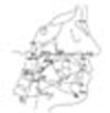 | Fig. 1Cephalometric landmarks. S (Sella), midpoint of the fossa hypophysealis; N (Nasion), anterior point at the frontonasal suture, Ba (Basion), most postero-inferior point on clivus; ANS (Anterior nasal spine), most anterior point of the nasal spine; PNS (Posterior nasal spine), most posterior point of the nasal spine; A, deepest anterior point in the concavity of the anterior maxilla; B, deepest anterior point in the concavity of the anterior mandible; Cd (Condylion), most postero-superior point of the condylar head; Gn (Gnathion), most antero-inferior point of the chin bone; Go (Gonion), a mid-point at the gonial angle located by bisecting the posterior and inferior borders of the mandible; Me (Menton), most inferior point of the chin bone; P, most inferior tip of soft palate; H, most antero-superior point of the hyoid bone; V, most antero-inferior point of the epiglottic fold; TT, most anterior point of the tip of the tongue; C3, most antero-inferior point of the third cervial vertebrae; MP (Mandibular plane), a tangent line constructed from Me to mandibular inferior border.
|
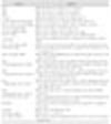

Table 3
Cephalometric measurements in simple snorers and OSA (obstructive sleep apnea syndrom) patients according
to obesity
![]()
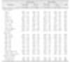
Table 4
Correlations between cephalometric measurements and AHI (apnea-hypopnea index) in OSA patients

![]()

References
1. Guilleminault C, Tilkian A, Dement WC. The sleep apnea syndromes. Annu Rev Med. 1976. 27:465–484.

2. American Academy of Sleep Medicine Task Force. Sleep-related breathing disorders in adults: recommendations for syndrome definition and measurement techniques in clinical research. The Report of an American Academy of Sleep Medicine Task Force. Sleep. 1999. 22:667–689.
3. Hoffstein V, Mateika S. Differences in abdominal and neck circumferences in patients with and without obstructive sleep apnoea. Eur Respir J. 1992. 5:377–381.
4. Schmidt-Nowara W, Lowe A, Wiegand L, Cartwright R, Perez-Guerra F, Menn S. Oral appliances for the treatment of snoring and obstructive sleep apnea: a review. Sleep. 1995. 18:501–510.
5. Young T, Palta M, Dempsey J, Skatrud J, Weber S, Badr S. The occurrence of sleep-disordered breathing among middleaged adults. N Engl J Med. 1993. 328:1230–1235.
6. Kripke DF, Ancoli-Israel S, Klauber MR, Wingard DL, Mason WJ, Mullaney DJ. Prevalence of sleep-disordered breathing in ages 40-64 years: a population-based survey. Sleep. 1997. 20:65–76.
7. Bixler EO, Vgontzas AN, Lin HM, Ten Have T, Rein J, Vela-Bueno A, et al. Prevalence of sleep-disordered breathing in women: effects of gender. Am J Respir Crit Care Med. 2001. 163:608–613.
8. Kim J, In K, Kim J, You S, Kang K, Shim J, et al. Prevalence of sleep-disordered breathing in middle-aged Korean men and women. Am J Respir Crit Care Med. 2004. 170:1108–1113.
9. Practice parameters. Polysomnography task force, American sleep disorders association standards of practice committee. Sleep. 1997. 20:406–422.
10. Haraldsson PO, Carenfelt C, Diderichsen F, Nygren A, Tingvall C. Clinical Symptoms of sleep apnea syndrome and automobile accidents. ORL J Otorhinolaryngol Relat Spec. 1990. 52:57–62.
11. George CF, Smiley A. Sleep apnea & automobile crashes. Sleep. 1999. 22:790–795.
12. Parish JM, Somers VK. Obstructive sleep apnea and cardiovascular disease. Mayo Clin Proc. 2004. 79:1036–1046.
13. Riley R, Guilleminault C, Herran J, Powell N. Cephalometric analysis and flow-volume loops in obstructive sleep apnea patients. Sleep. 1983. 6:303–311.
14. Tangugsorn V, Krogstad O, Espeland L, Lyberg T. Obstructive sleep apnea: a canonical correlation of cephalometric and selected demographic variables in obese and nonobese patients. Angle Orthod. 2001. 71:23–35.
15. Ono T, Lowe AA, Ferguson KA, Fleetham JA. Associations among upper airway structure, body position, and obesity in skeletal Class I male patients with obstructive sleep apnea. Am J Orthod Dentofacial Orthop. 1996. 109:625–634.
16. deBerry-Borowiecki B, Kukwa A, Blanks RH. Cephalometric analysis for diagnosis and treatment of obstructive sleep apnea. Laryngoscope. 1988. 98:226–234.
17. Partinen M, Guilleminault C, Quera-Salva MA, Jamieson A. Obstructive sleep apnea and cephalometric roentgenograms. The role of anatomic upper airway abnormalities in the definition of abnormal breathing during sleep. Chest. 1988. 93:1199–1205.
18. Baik UB, Suzuki M, Ikeda K, Sugawara J, Mitani H. Relationship between cephalometric characteristics and obstructive sites in obstructive sleep apnea syndrome. Angle Orthod. 2002. 72:124–134.
19. Guilleminault C, Quera-Salva MA, Partinen M, Jamieson A. Women and the obstructive sleep apnea syndrome. Chest. 1988. 93:104–109.
20. Ong KC, Clerk AA. Comparison of the severity of sleep disordered breathing in Asian and Caucasian patients seen at a sleep disorders center. Respir Med. 1998. 92:843–848.
21. Li KK, Powell NB, Kushida C, Riley RW, Adornato B, Guilleminault C. A comparison of Asian and white patients with obstructive sleep apnea syndrome. Laryngoscope. 1999. 109:1937–1940.
22. Liu Y, Lowe AA, Zeng X, Fu M, Fleetham JA. Cephalometric comparisons between Chinese and Caucasian patients with obstructive sleep apnea. Am J Orthod Dentofacial Orthop. 2000. 117:479–485.
23. Horner RL, Mohiaddin RH, Lowell DG, Shea SA, Burman ED, Longmore DB, et al. Sites and sizes of fat deposits around the pharynx in obese patients with obstructive sleep apnoea and weight matched controls. Eur Respir J. 1989. 2:613–622.
24. Davies RJ, Stradling JR. The relationship between neck circumference, radiographic pharyngeal anatomy, and the obstructive sleep apnoea syndrome. Eur Respir J. 1990. 3:509–514.
25. Deegan PC, McNicholas WT. Predictive value of clinical features for the obstructive sleep apnea syndrome. Eur Respir J. 1996. 9:117–124.
26. Rubinstein I, Colapinto N, Rotstein LE, Brown IG, Hoffstein V. Improvement in upper airway function after weight loss in patients with obstructive sleep apnea. Am Rev Respir Dis. 1988. 138:1192–1195.
27. Schwartz AR, Gold AR, Schubert N, Stryzak A, Wise RA, Permutt S, et al. Effect of weight loss on upper airway collapsibility in obstructive sleep apnea. Am Rev Respir Dis. 1991. 144:494–498.
28. World Health Organization Western Pacific Region. International Association for the Study of Obesity. International Obesity Task Force. The Asian-Pacific perspective: redefining obesity and its treatment. 2000. Geneva, Switzerland: WHO Western Pacific Region.
29. Li KK, Kushida C, Powell NB, Riley RW, Guilleminault C. Obstructive sleep apnea syndrome: a comparison between Far-East Asian and white men. Laryngoscope. 2000. 110:1689–1693.
30. Tsuchiya M, Lowe AA, Pae EK, Fleetham JA. Obstructive sleep apnea subtypes by cluster analysis. Am J Orthod Dentofacial Orthop. 1992. 101:533–542.
31. Hui DS, Ko FW, Chu AS, Fok JP, Chan MC, Li TS, et al. Cephalometric assessment of craniofacial morphology in Chinese patients with obstructive sleep apnoea. Respir Med. 2003. 97:640–646.
32. Yu X, Fujimoto K, Urushibata K, Matsuzawa Y, Kubo K. Cephalometric analysis in obese and nonobese patients with obstructive sleep apnea syndrome. Chest. 2003. 124:212–218.
33. Miles PG, Vig PS, Weyant RJ, Forrest TD, Rockette HE Jr. Craniofacial structure and obstructive sleep apnea syndrome-a qualitative analysis and meta-analysis of the literature. Am J Orthod Dentofacial Orthop. 1996. 109:163–172.
34. Wilms D, Popovich J, Conway W, Fujita S, Zorick F. Anatomic abnormalities in obstructive sleep apnea. Ann Otol Rhinol Laryngol. 1982. 91:595–596.
35. Finkelstein Y, Wexler D, Horowitz E, Berger G, Nachmani A, Shapiro-Feinberg M, et al. Frontal and lateral cephalometry in patients with sleep-disordered breathing. Laryngoscope. 2001. 111:634–641.
1. Pracharktam N, Hans MG, Strohl KP, Redline S. Upright and supine cephalometric evaluation of obstructive sleep apnea syndrome and snoring subjects. Angle Orthod. 1994. 64:63–73.
37. Kong HW, Lee HJ, Choi YS, Rha JH, Ha CK, Hwangc DU, et al. Clinical predictors of obstructive sleep apnea. J Korean Neurol Assoc. 2005. 23:324–329.
38. Otsuka R, Almeida FR, Lowe AA, Ryan F. A comparison of responders and nonresponders to oral appliance therapy for the treatment of obstructive sleep apnea. Am J Orthod Dentofacial Orthop. 2006. 129:222–229.
39. Littner M, Hirshkowitz M, Davila D, Anderson WM, Kushida CA, Woodson BT, et al. Practice parameters for the use of auto-titrating continuous positive airway pressure devices for titrating pressures and treating adult patients with obstructive sleep apnea syndrome. An American Academy of Sleep Medicine report. Sleep. 2002. 25:143–147.
40. Fujita S, Conway W, Zorick F, Roth T. Surgical correction of anatomic abnormalities in obstructive sleep apnea syndrome: uvulopalatopharyngoplasty. Otolaryngol Head Neck Surg. 1981. 89:923–934.
41. American Sleep Disorders Association. Practice parameters for the treatment of snoring and obstructive sleep apnea with oral appliances. American Sleep Disorders Association. Sleep. 1995. 18:511–513.
 XML Download