

 PDF
PDF ePub
ePub Citation
 Print
Print


Abstract
Objective
To evaluate the discrepancies between initial STO and final STO in Class III malocclusions and to find which factors are related to the discrepancies.
Methods
Twenty patients were selected for the extraction group and 20 patients for the non-extraction group. They were diagnosed as skeletal Class III and received presurgical orthodontic treatment and mandibular set-back surgery at Pusan National University Hospital. The lateral cephalograms were analyzed for initial STO (T1s) at pretreatment and final STO (T2s) after presurgical orthodontic treatment, and specified the landmarks as coordinates of the X and Y axes.
Results
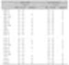
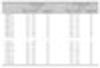
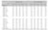
Differences in hard
tissue points (T1s-T2s) in the X coordinates of upper central incisor edge, upper first molar mesial end surface, lower central incisor apex, lower first molar mesial end surface and mesio-buccal cusp and Y coordinates of upper central incisor edge, upper central incisor apex, upper first molar mesio-buccal cusp were statistically significant in the extraction group. Differences in hard tissue points (T1s-T2s) in the X coordinates of upper central incisor edge, lower central incisor apex, lower first molar mesial end surface and Y coordinates of lower central incisor apex were statistically significant in the non-extraction group. In the extraction group, the upper arch length discrepancy (UALD) had a statistically significant effect on maxillary incisor and first molar estimation. Lower arch length discrepancy and IMPA had statistically significant effects on mandibular incisor estimation in both groups.
Figures and Tables
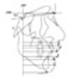
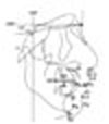 | Fig. 1Hard tissue and soft tissue landmarks. VRP, vertical reference plane; HRP, horizontal reference plane; S, sella; Na, nasion; A, subspinale; B, supramentale; Pg, pogonion; Me, menton; U1e, upper central incisor edge; U1a, upper central incisor apex; U6m, upper 1st molar mesial end surface; U6mbc, upper 1st molar mesio-buccal cusp tip; L1e, lower central incisor edge; L1a, lower central incisor apex; L6m, lower 1st molar mesial end surface; L6mbc, lower 1st molar mesio-buccal cusp tip; Sn, subnasale; SLS, superior labial sulcus; ILS, labrale superius; Stm, stomion; LI, labrale inferius; LS, inferior labial sulcus; Pg', soft tissue pogonion; Me', soft tissue menton.
|
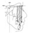 | Fig. 2Horizontal measurements of hard tissue landmarks. 1, VRP-A; 2, VRP-U1a; 3, VRP-U6m; 4, VRP-U6mbc; 5, VRP-L6mbc; 6, VRP-L6m; 7, VRP-L1e; 8, VRP-U1e; 9, VRP-L1a; 10, VRP-B; 11, VRP-Pg; 12, VRP-Me.
|
 | Fig. 3Horizontal measurements of hard tissue landmarks. 1, VRP-A; 2, VRP-U1a; 3, VRP-U6m; 4, VRP-U6mbc; 5, VRP-L6mbc; 6, VRP-L6m; 7, VRP-L1e; 8, VRP-U1e; 9, VRP-L1a; 10, VRP-B; 11, VRP-Pg; 12, VRP-Me.
|
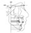
 | Fig. 4Vertical measurements of soft tissue landmarks. 1, HRP-Sn; 2, HRP-SLS; 3, HRP-LS; 4, HRP-Stm; 5, HRP-LI; 6, HRP-ILS; 7, HRP-Pg'; 8, HRP-Me'.
|
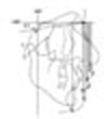
 | Fig. 5Horizontal measurements of soft tissue landmarks. 1, HRP-Sn; 2, HRP-SLS; 3, HRP-LS; 4, HRP-Stm; 5, HRP-LI; 6, HRP-ILS; 7, HRP-Pg'; 8, HRP-Me'.
|
References
1. Vig KD, Ellis E 3rd. Diagnosis and treatment planning for the surgical-orthodontic patient. Dent Clin North Am. 1990. 34:361–384.
2. Tompach PC, Wheeler JJ, Fridrich KL. Orthodontic considerations in orthognathic surgery. Int J Adult Orthodon Orthognath Surg. 1995. 10:97–107.
3. Proffit WR, White RP. Surgical orthodontic treatment. 1991. St Louis: Mosby;202–215.
4. Park JH, Hwang CJ. A study on the preoperative prediction values versus the postoperative actual values in Class III two jaw surgery patients. J Korean Assoc Maxillofac Plast Reconstr Surg. 2003. 25:238–248.
5. Choi YS, Son WS. A comparative study on the postsurgical changes between one jaw surgery and two-jaw surgery in skeletal Class III patients. Korean J Orthod. 1997. 27:297–313.
6. Choi YK, Suhr CH. Hard and soft tissue changes after orthognathic surgery of mandibular prognathism. Korean J Orthod. 1993. 23:707–724.
7. Kim JR, Kim TK, Chung IK, Yang DK, Park SB, Son WS, et al. Cephalometric analysis of postsurgical behavior of mandibular prognathism. J Korean Assoc Maxillofac Plast Reconstr Surg. 1993. 15:123–128.
8. Lee HS, Park YC. A cephalometric study of profile changes following orthognathic surgery in patients with mandibular prognathism. Korean J Orthod. 1987. 17:299–310.
9. Arnett GW, Gunson MJ. Facial planning for orthodontists and oral surgeons. Am J Orthod Dentofacial Orthop. 2004. 126:290–295.

10. Arnett GW, Jelic JS, Kim J, Cummings DR, Beress A, Worley CM Jr, et al. Soft tissue cephalometric analysis: Diagnosis and treatment planning of dentofacial deformity. Am J Orthod Dentofacial Orthop. 1999. 116:239–253.
11. McNeill RW, Proffit WR, White RP. Cephalometric prediction for orthodontic surgery. Angle Orthod. 1972. 42:154–164.
12. Lines PA, Steinhauser EW. Soft tissue changes in relationship to movement of hard structures in orthognathic surgery: a preliminary report. J Oral Surg. 1974. 32:891–896.
13. Robinson SW, Speidel TM, Isaacson RJ, Worms FW. Soft tissue profile change produced by reduction of mandibular prognathism. Angle Orthod. 1972. 42:227–235.
14. Worms FW, Isaacson RJ, Speidel TM. Surgical orthodontic treatment planning: Profile analysis and mandibular surgery. Angle Orthod. 1976. 46:1–25.
15. Gossett CB, Preston CB, Dunford R, Lampasso J. Prediction accuracy of computer-assisted surgical visual treatment objectives as compared with conventional visual treatment objectives. J Oral Maxillofac Surg. 2005. 63:609–617.
16. Eckhardt CE, Cunningham SJ. How predictable is orthognathic surgery? Eur J Orthod. 2004. 26:303–309.
17. Friede H, Kahnberg KE, Adell R, Ridell A. Accuracy of cephalometric prediction in orthognathic surgery. J Oral Maxillofac Surg. 1987. 45:754–760.
18. Kim NK, Lee C, Kang SH, Park JW, Kim MJ, Chang YI. A three-dimensional analysis of soft and hard tissue changes after a mandibular setback surgery. Comput Methods Programs Biomed. 2006. 83:178–187.
19. Bryan DC, Hunt NP. Surgical accuracy in orthognathic surgery. Br J Oral Maxillofac Surg. 1993. 31:343–349.
20. Mankad B, Cisneros GJ, Freeman K, Eisig SB. Prediction accuracy of soft tissue profile in orthognathic surgery. Int J Adult Orthodon Orthognath Surg. 1999. 14:19–26.
21. Yang WS, Baik HS. A study on the extracellular matrix in the artificially created cleft lip wound healing of rabbit fetuses. Korean J Orthod. 1998. 28:865–875.
22. Pospisil OA. Reliability and feasibility of prediction tracing in orthognathic surgery. J Craniomaxillofac Surg. 1987. 15:79–83.
23. Lee SJ, Hong SJ, Kim YH, Baek SH, Suhr CH. Effect of maxillary premolar extracion on transverse arch dimension in Class III surgical-orthodontic treatment. Korean J Orthod. 2005. 35:23–34.
24. Tweed CH. The Frankfort-mandibular incisor angle in orthodontic diagnosis, treatment planning and prognosis. Angle Orthod (FMIA). 1954. 24:121–169.
25. Hixon EH. Cephalometrics: A perspective. Angle Orthod. 1972. 42:200–211.
26. Steiner CC. The use of cephalometrics as an aid to planning and assessing orthodontic treatment. Am J Orthod. 1960. 46:721–735.
27. Choi BT. Steps of preparation for orthognathic surgery. 2004. Seoul: JeeSung Publishing.
28. Yang SD. Surgical treatment objectives. J Korean Dent Assoc. 2007. 45:404–413.
29. Hwang CJ, Moon JL. The limitation of alveolar bone remodeling during retraction of the upper anterior teeth. Korean J Orthod. 2001. 31:97–105.
30. Chang IH, Lee YJ, Park YG. A comparative study of soft tissue changes with mandibular one jaw surgery and double jaw surgery in Class III malocclusion. Korean J Orthod. 2006. 36:63–73.
31. Cho EJ, Yang WS. Soft tissue changes after double jaw surgery in skeletal Class III maloccluaion. Korean J Orthod. 1996. 26:1–16.
32. Wolford LM, Hilliard FW, Dugan DJ. Surgical treatment objective: a systemic approach to the prediction tracing. 1985. St. Louis: Mosby;54–74.
33. Kwon MJ, Baik HS, Lee WY. A study on the accuracy of profile change prediction by video imaging (Power Ceph® Ver 3.3) in Class III two jaw surgery patients. Korean J Orthod. 1999. 29:285–301.
34. Sinclair PM, Kilpelainen P, Phillips C, White RP Jr, Rogers L, Sarver DM. The accuracy of video imaging in orthognathic surgery. Am J Orthod Dentofacial Orthop. 1995. 107:177–185.
35. Jacobs JD, Sinclair PM. Principles of orthodontic mechanics in orthognathic surgery cases. Am J Orthod. 1983. 84:399–407.
36. Kim SJ, Park SY, Woo HH, Park EJ, Kim YH, Lee SJ, et al. A study on the limit of orthodontic treatment. Korean J Orthod. 2004. 34:165–175.
37. Hwang CJ, Kwon HJ. A study on the preorthodotic prediction values versus the actual postorthodontic values in Class III surgery patients. Korean J Orthod. 2003. 33:1–9.
38. Chang JH, Lee SJ, Kim TW. Evaluation of nasolabial angle in adult patients with skeletal Class III malocclusion. Korean J Orthod. 2007. 37:272–282.
39. Kobayashi T, Watanabe I, Ueda K, Nakajima T. Stability of the mandible after sagittal ramus osteotomy for correction of prognathism. J Oral Maxillofac Surg. 1986. 44:693–697.
40. Lee RT. The benefits of post-surgical orthodontic treatment. Br J Orthod. 1994. 21:265–274.
 XML Download