

 PDF
PDF ePub
ePub Citation
 Print
Print


Abstract
Objective
The purpose of this study was to investigate the stability of mini-implants in relation to loading time.
Methods
A total of 48 mini-implants (ORLUS, Ortholution, Korea) were placed into the buccal alveolar bone of the mandible in 8 male beagle dogs. Orthodontic force (200 - 250 gm) was applied immediately for the immediate loading group while force application was delayed for 3 weeks in the delayed loading group. For the subsequent loading periods (3, 6, 12 weeks), BIC (bone implant contact) and BV/TV (bone volume/total volume) and mobility test were carried out.
Results
The immediate loading group showed no changes in BIC from 3 to 12 weeks, while the delayed loading group showed a significant increase in BIC between 3 and 12 weeks (p < 0.05). The BV/TV of the delayed loading group significantly increased from 6 to 12 weeks of loading (p < 0.05), while the BV/TV of the immediate loading group decreased from 3 to 12 weeks of loading. However, there was no significant difference in BV/TV between experimental groups. The mobility of the immediate loading group was not significantly different from that of the delayed loading group after 12 weeks of loading (p < 0.05).
Figures and Tables
Fig. 1
Implantation sites of orthodontic mini-implants (A) and intra-oral photo (B). Orthodontic forces to both immediate and delayed groups were loaded reciprocally with elastic chain.
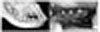
Fig. 2
Timetable for installation of mini-implants. W, weeks; DL, delayed loading group; IL, immediate loading group; HC, healing control group.
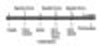
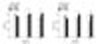
Fig. 3
Subsequent photographs of mini-implants for each group after installation. Histological section showed well-organized vital bone in contact with the implant after loading. Mature lamellar bone was visible at 6 weeks. Toluidine blue; × 100; 3 w, 3 week; 6 w, 6 weeks; 12 w, 12 weeks; scale bar shows 100 um.
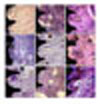
Fig. 4
Histological findings at week 6 of the immediate loading group. Intimate bone-implant contact was observed (left, × 100); osteoclast-like cell was shown at implant-bone surface (center) and bone marrow (right, × 200); B, alveolar bone; M, mini-implant.
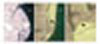
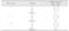
Fig. 5
Bone implant contact (BIC, %) and bone volume/total volume (BV/TV, %) of each group for different time periods. *p < 0.05. IL, Immediate loading group; DL, delayed loading group; HC, healing control group.
References
1. Kanomi R. Mini-implant for orthodontic anchorage. J Clin Orthod. 1997. 31:763–767.
2. Park HS, Bae SM, Kyung HM, Sung JH. Micro-implant anchorage for treatment of skeletal Class I bialveolar protrusion. J Clin Orthod. 2001. 35:417–422.
3. Kyung SH, Choi JH, Park YC. Miniscrew anchorage used to protract lower second molars into first molar extraction sites. J Clin Orthod. 2003. 37:575–579.
4. Lee JS, Kim DH, Park YC, Kyung SH, Kim TK. The efficient use of midpalatal miniscrew implants. Angle Orthod. 2004. 74:711–714.
5. Roth A, Yildirim M, Diedrich P. Forced eruption with microscrew anchorage for preprosthetic leveling of the gingival margin. Case report. J Orofac Orthop. 2004. 65:513–519.

6. Kyung SH, Choi HW, Kim KH, Park YC. Bonding orthodontic attachments to miniscrew heads. J Clin Orthod. 2005. 39:348–353. quiz 369.
7. Park HS, Lee SK, Kwon OW. Group distal movement of teeth using microscrew implant anchorage. Angle Orthod. 2005. 75:602–609.
8. Kim HJ, Yun HS, Park HD, Kim DH, Park YC. Soft-tissue and cortical-bone thickness at orthodontic implant sites. Am J Orthod Dentofacial Orthop. 2006. 130:177–182.
9. Turley PK, Kean C, Schur J, Stefanac J, Gray J, Hennes J, et al. Orthodontic force application to titanium endosseous implants. Angle Orthod. 1988. 58:151–162.
10. Roberts WE, Helm FR, Marshall KJ, Gongloff RK. Rigid endosseous implants for orthodontic and orthopedic anchorage. Angle Orthod. 1989. 59:247–256.
11. Wehrbein H, Diedrich P. Endosseous titanium implants during and after orthodontic load--an experimental study in the dog. Clin Oral Implants Res. 1993. 4:76–82.
12. Roberts WE, Nelson CL, Goodacre CJ. Rigid implant anchorage to close a mandibular first molar extraction site. J Clin Orthod. 1994. 28:693–704.
13. Akin-Nergiz N, Nergiz I, Schulz A, Arpak N, Niedermeier W. Reactions of peri-implant tissues to continuous loading of osseointegrated implants. Am J Orthod Dentofacial Orthop. 1998. 114:292–298.
14. Melsen B, Costa A. Immediate loading of implants used for orthodontic anchorage. Clin Orthod Res. 2000. 3:23–28.
15. Deguchi T, Takano-Yamamoto T, Kanomi R, Hartsfield JK Jr, Roberts WE, Garetto LP. The use of small titanium screws for orthodontic anchorage. J Dent Res. 2003. 82:377–381.
16. Miyawaki S, Koyama I, Inoue M, Mishima K, Sugahara T, Takano-Yamamoto T. Factors associated with the stability of titanium screws placed in the posterior region for orthodontic anchorage. Am J Orthod Dentofacial Orthop. 2003. 124:373–378.
17. Cheng SJ, Tseng IY, Lee JJ, Kok SH. A prospective study of the risk factors associated with failure of mini-implants used for orthodontic anchorage. Int J Oral Maxillofac Implants. 2004. 19:100–106.
18. Park HS, Jeong SH, Kwon OW. Factors affecting the clinical success of screw implants used as orthodontic anchorage. Am J Orthod Dentofacial Orthop. 2006. 130:18–25.
19. Park HS, Yen S, Jeoung SH. Histologic and biomechanical characteristics of orthodontic self-drilling and self-tapping microscrew implants. Korean J Orthod. 2006. 36:295–397.
20. Kim JW, Ahn SJ, Chang YI. Histomorphometric and mechanical analyses of the drill-free screw as orthodontic anchorage. Am J Orthod Dentofacial Orthop. 2005. 128:190–194.
21. Frost HM. Perspectives: bone's mechanical usage windows. Bone Miner. 1992. 19:257–271.
22. Freire JN, Silva NR, Gil JN, Magini RS, Coelho PG. Histomorphologic and histomophometric evaluation of immediately and early loaded mini-implants for orthodontic anchorage. Am J Orthod Dentofacial Orthop. 2007. 131:704. e1-9.
23. Gray JB, Steen ME, King GJ, Clark AE. Studies on the efficacy of implants as orthodontic anchorage. Am J Orthod. 1983. 83:311–317.
24. Mori S, Burr DB. Increased intracortical remodeling following fatigue damage. Bone. 1993. 14:103–109.
25. Isidor F. Mobility assessment with the Periotest system in relation to histologic findings of oral implants. Int J Oral Maxillofac Implants. 1998. 13:377–383.
26. Schulte W, Lukas D. The Periotest method. Int Dent J. 1992. 42:433–440.
27. Tricio J, Laohapand P, van Steenberghe D, Quirynen M, Naert I. Mechanical state assessment of the implant-bone continuum: a better understanding of the Periotest method. Int J Oral Maxillofac Implants. 1995. 10:43–49.
28. Trisi P, Rao W, Rebaudi A. A histometric comparison of smooth and rough titanium implants in human low-density jawbone. Int J Oral Maxillofac Implants. 1999. 14:689–698.
29. Oyonarte R, Pilliar RM, Deporter D, Woodside DG. Peri-implant bone response to orthodontic loading: Part 2. Implant surface geometry and its effect on regional bone remodeling. Am J Orthod Dentofacial Orthop. 2005. 128:182–189.
 XML Download