

 PDF
PDF ePub
ePub Citation
 Print
Print


Abstract
Objective
The purpose of this study was to evaluate the upper airway dimensional change according to maxillary superior movement after orthognathic surgery and to identify the relationship between the amount of maxillary movement and upper airway dimensional changes.
Methods
The samples consisted of 24 adult patients (9 males and 15 females) who had a skeletal discrepancy and had received presurgical orthodontic treatment. They underwent Le Fort I superior impaction osteotomy and mandibular setback surgery. Cephalometric x-rays were taken at 3 stages - T0 (before orthognathic surgery), T1 (just or within 2 weeks after orthognathic surgery), T2 (6 months after surgery)
Results
1, Pharyngeal airway space (PAS (R)-nasopharynx) was decreased after surgery (T1) but recovered at 6 months after surgery; 2, Pharyngeal airway space (PAS (NL)-palatal plane) was increased after surgery and at 6 months after surgery; 3, Pharyngeal airway space (PAS (OL)-occlusal plane) was increased at T1 and was decreased at T2; 4, Soft palate thickness was increased at T1 but it became the same or thinner at T2; 5, There is no statistically significant relation between the amount of maxillary superior movement and pharyngeal airway space.
Figures and Tables


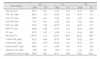
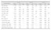
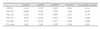
Table 6
Changes in Vertical-PNS, ANS and Horizontal-PNS, ANS, B point - S-perpendicular plane after surgery

![]()

References
1. Park JW, Lee ET, Min SK, Lee DK. Stability of two-jaw surgery and single mandibular surgery in mandibular prognathism. J Korean Assoc Maxillofac Plast Recontr Surg. 2002. 24:406–414.
2. Jeong MH, Choi JH, Kim BH, Kim SG, Nahm DS. Soft tissue changes after double jaw rotation surgery in skeletal Class III malocclusion. J Korean Oral Maxillofac Surg. 2006. 32:559–565.
3. Bell WH, Jacobs JD, Quejada JG. Simultaneous repositioning of the maxilla, mandible, and chin. Treatment planning and analysis of soft tissues. Am J Orthod. 1986. 89:28–50.

4. Komori E, Aigase K, Sugisaki M, Tanabe H. Cause of early skeletal relapse after mandibular setback. Am J Orthod Dentofacial Orthop. 1989. 95:29–36.
5. Pepersack WJ, Chausse JM. Long-term follow up of the sagittal splitting technique for correction of mandibular prognathism. J Maxillofac Surg. 1978. 6:117–140.
6. Mommaerts MY, Hadjianghelou O. Positional changes after mandibular advancement by sagittal split osteotomies and wire osteosynthesis. J Craniomaxillofac Surg. 1990. 18:93–106.
7. Solow B, Siersbaek-Nielsen S, Greve E. Airway adequacy, head posture, and craniofacial morphology. Am J Orthod. 1984. 86:214–223.
8. Joseph AA, Elbaum J, Cisneros GJ, Eisig SB. A cephalometric comparative study of the soft tissue airway dimensions in persons with hyperdivergent and normodivergent facial patterns. J Oral Maxillofac Surg. 1998. 56:135–139.
9. Dunn GF, Green LJ, Cunat JJ. Relationships between variation of mandibular morphology and variation of nasopharyngeal airway size in monozygotic twins. Angle Orthod. 1973. 43:129–135.
10. Lubart J. The adenoid problem. Arch Pediatr. 1960. 77:491–495.
11. Handelman CS, Osborne G. Growth of the nasopharynx and adenoid development from one to eighteen years. Angle Orthod. 1976. 46:243–259.
12. Robin R. Effect of nasal airway obstruction on facial growth. Ear Nose Throat J. 1987. 66:212–219.
13. Ahn SC, Suhr CH. A study on nasal respiratory patency in the growing children with anterior crossbite. Korean J Orthod. 1992. 22:179–203.
14. Chin KS, Shon WS. The relationships between the postoperative stability and the changes in the tongue position, the hyoid bone position and the upper airway size after orthognathic surgery in patients with mandibular prognathism. Korean J Orthod. 1993. 23:693–705.
15. Athanasiou AE, Toutountzakis N, Mavreas D, Ritzau M, Wenzel A. Alterations of hyoid bone position and pharyngeal depth and their relationship after surgical correction of mandibular prognathism. Am J Orthod Dentofacial Orthop. 1991. 100:259–265.
16. Greco JM, Frohberg U, Van Sickels JE. Long-term airway space changes after mandibular setback using bilateral sagittal split osteotomy. Int J Oral Maxillofac Surg. 1990. 19:103–105.
17. Cho SJ, Kim YG. A study on relation of position of hyoid bone and upper airway dimensional change according to chin movement in persons with skeletal Class III facial pattern after orthognathic surgery. J Korean Assoc Maxillofac Plast Recontr Surg. 2000. 22:343–350.
18. Bae JS, Kim KH, Park HS, Huh JK, Park KH. Cephalometric study of posterior airway space and hyoid bone position in patients affected by Class II malocclusion and treated with orthognathic surgery. J Korean Assoc Maxillofac Plast Recontr Surg. 2001. 23:540–552.
19. Chen F, Terada K, Hua Y, Saito I. Effects of bimaxillary surgery and mandibular setback surgery on pharyngeal airway measurements in patients with Class III skeletal deformities. Am J Orthod Dentofacial Orthop. 2007. 131:372–377.
20. Epker BN, Turvey T, Fish LC. Indication for simultaneous mobilization of the maxilla and mandible for the correction of dentofacial deformities. Oral Surg Oral Med Oral Pathol. 1982. 54:369–381.
21. Reyneke JP, Bryant RS, Suuronen R, Becker PJ. Postoperative skeletal stability following clockwise and counter-clockwise rotation of the maxillomandibular complex compared to conventional orthognathic treatment. Br J Oral Maxillofac Surg. 2007. 45:56–64.
22. Lee YJ, Sohn BW. A study on the postoperative stability of occlusal plane in Class III orthognathic surgery patients. Korean J Orthod. 2000. 30:643–655.
23. Hochban W, Schürmann R, Brandenburg U, Conradt R. Mandibular setback for surgical correction of mandibular hyperplasia- does it provoke sleep disorder? Int J Oral Maxillofac Surg. 1996. 25:333–338.
24. Samman N, Tang SS, Xia J. Cephalometric study of the upper airway in surgically corrected Class III skeletal deformity. Int J Adult Orthodon Orthognath Surg. 2002. 17:180–190.
25. Cakarne D, Urtane I, Skagers A. Pharyngeal airway sagittal dimension in patients with Class III skeletal dentofacial deformity before and after bimaxillary surgery. Stomatologija. 2003. 5:13–16.
26. Proffit WR, White RP, Sarver DM. Contemporary treatment of dentofacial deformity. 2003. St Louis: Mosby;302.
27. Son WS, Choi YS. Evaluation of hyoid bone position and airway size in Class malocculsion. Korean J Orthod. 1996. 26:247–254.
28. Solow B, Kreiborg S. Soft tissue stretching: a possible control factor in craniofacial morphogenesis. Scan J Dent Res. 1977. 85:505–507.
29. Lowe AA, Fleetham JA, Adachi S, Ryan CF. Cephalometric and computed tomographic predictors of obstructive sleep apnea severity. Am J Orthod Dentofacial Orthop. 1995. 107:589–595.
30. Malkoc S, Usumez S, Nur M, Donaghy CE. Reproducibility of airway dimensions and tongue and hyoid positions on lateral cephalograms. Am J Orthod Dentofacial Orthop. 2005. 128:513–516.
31. Lee YS, Kim JC. A cephalometric study on the airway size according to the types of the malocclusion. Korean J Orthod. 1995. 25:19–29.
32. Tselnik M, Pogrel MA. Assessment of the pharyngeal airway space after mandibular setback surgery. J oral maxillofac Surg. 2000. 58:282–285.
33. Wenzel A, Williams S, Ritzau M. Changes in head posture and nasopharyngeal airway following surgical correction of mandibular prognathism. Eur J Orthod. 1989. 11:37–42.
34. Shin KY, Lee DK, Oh SH, Sung HM, Lee SH. Comparison of speech patterns according to the degree of surgical setback in mandibular prognathic patients. J Korean Assoc Maxillofac Plast Recontr Surg. 2001. 23:48–58.
35. Nam SY, Suh SB, Chang Y. The effect of tonsillectomy and adenoidectomy on nasality. Korean J Otolaryngol. 1999. 42:354–357.
36. Wickwire NA, White RP Jr, Proffit WR. The effect of mandibular osteotomy on tongue position. J Oral Surg. 1972. 30:184–190.
37. Chung DH, Lee KS. A study on changes of airway, tongue, and hyoid position following orthognathic surgery. Korean J Orthod. 1998. 28:487–498.
38. Turvey TA, Hall DJ, Warren DW. Alterations in nasal airway resistance following superior repositioning of the maxilla. Am J Orthod. 1984. 85:109–114.
39. Walker DA, Turvey TA, Warren DW. Alterations in Nasal Respiration and Nasal Airway Size Following Superior Repositioning of the Maxilla. J Oral Maxillofac Surg. 1988. 46:276–281.
 XML Download