

 PDF
PDF ePub
ePub Citation
 Print
Print


Abstract
Purpose
The purpose of this study was to assess the hard and soft tissue changes associated with mandibular bilateral sagittal split osteotomy and genioplasty.
Methods
This is a retrospective study of 40 patients who underwent either bilateral sagittal split osteotomy for mandibular setback (BSSO group, n = 20) or in combination with advancement genioplasty (Genio group, n = 20). Lateral radiographs, were taken before and immediately after surgery, and at least 6 months after surgery.
Results
Comparing hard and soft tissue changes between the BSSO group and Genio group, there were significant differences in the lower incisor, soft tissue B point (B'), and soft tissue Pogonion (Pg') (p < 0.5). The mean ratio of hard and soft tissue changes for B/B', Pg/Pg', and Menton/soft tissue Menton after surgery in the BSSO group was 0.997, 0.965, and 1.022 respectively, and 0.824, 0.602, and 0.887 respectively in the genio group. Significant differences were found between the two groups. There were significant differences in lip thickness (B-B', Pg-Pg') in the Genioplasty group between pre and postsurgery, but not in the BSSO group. Pogonion to Labrale inferior and B' had a correlation coefficient of 0.833, 0.922, respectively for the BSSO group, and 0.775, 0.799 for the Genio group.
Conclusions
The results indicate that there is a significant difference between bilateral sagittal split osteotomy with or without genioplasty in the lower facial esthetics values. The combination of mandibular setback and genioplasty had a smaller change in soft tissue thickness of the symphysis area after surgery than that of mandibular setback only.
Figures and Tables
Fig 1
Cephalometric landmarks of hard and soft tissue points and associated reference planes. 1, U1 ; 2, L1; 3, infra; 4, B; 5, Pg; 6, Me; 7, Stos; 8, Stoi; 9, Li; 10, B'; 11, Pg'; 12, Me'; 13, S; 14, Na.
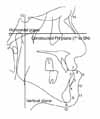
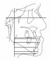
Fig 4
Vertical measurements of hard tissue landmarks. 13, U1; 14, L1; 15, infra; 16, B; 17, Pg; 18, Me.
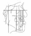
Fig 5
Vertical measurements of soft tissue landmarks. 19, Stos; 20, Stoi; 21, Li; 22, B'; 23, Pg'; 24, Me'.
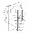
Fig 6
Lip thickness measurements. 25, U1-Li; 26, L1-Li; 27, Infra-B'; 28, B-B'; 29, Pg-Pg'; 30, U1-Stoi; 31, L1-Stoi.
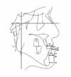
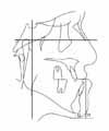
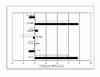
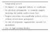
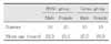
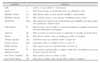
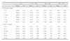
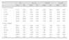
Table 7
Horizontal changes of Hard and soft tissue landmarks between each group (T1 - T3) (unit: mm)
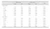
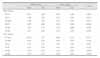
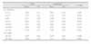
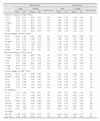
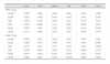
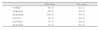
Table 11
Correlation coefficients between hard tissue and soft tissue horizontal changes for each group (T1 - T3)

References
1. Stricker G, Clifford E, Cohen LK, Giddon DB, Meskin LH, Evans CA. Psychosocial aspects of craniofacial disfigurement. Am J Orthod. 1979. 76:410–422.

2. Ewing M, Ross RB. Soft tissue response to mandibular advancement and genioplasty. Am J Orthod Dentofacial Orthop. 1992. 101:550–555.
3. Ellis E 3rd, Dechow PC, McNamara JA Jr, Carlson DS, Liskiewicz WE. Advancement genioplasty with and without soft tissue pedicle; An experimental investigation. J Oral Maxillofac Surg. 1984. 42:637–645.
4. Heiple KG, Chase SW, Herndon CH. A comparative study of the healing process following different types of bone transplantation. J Bone Joint Surg Am. 1963. 45:1593–1616.
5. Thompson N, Casson J. Experimental onlay bone grafts to the jaws. A preliminary study in dogs. Plast Reconstr Surg. 1970. 46:341–349.
6. Dann JJ, Epker BN. Proplast genioplasty; a retrospective study with treatment recommendations. Angle Orthod. 1977. 47:173–185.
7. Friedland JA, Coccaro PJ, Converse JM. Retrospective cephalometric analysis of mandibular bone absorption under silicone rubber chin implants. Plast Reconstr Surg. 1976. 57:144–151.
8. Robinson M, Shuken R. Bone resorption under plastic chin implants. J Oral Surg. 1969. 27:116–118.
9. Peled IJ, Wexler MR, Ticher S, Lax EE. Mandibular resorption from silicone chin implants in children. J Oral Maxillofac Surg. 1986. 44:346–348.
10. Park HS, Ellis E 3rd, Fonseca RJ, Reynolds ST, Mayo KH. A retrospective study of advancement genioplasty. Oral Surg Oral Med Oral Pathol. 1989. 67:481–489.
11. Björk N Eliasson S, Wictorin L. Changes of facial profile after surgical treatment of mandibular protrusion. Scand J Plast Reconstr Surg. 1971. 5:41–46.
12. Fromm B, Lundberg M. The soft tissue facial profile before and after surgical correction of mandibular protrusion. Acta Odontol Scand. 1970. 28:157–177.
13. Robinson SW, Spiedel TM, Isaacson RJ, Worms FW. Soft tissue profile change produced by reduction of mandibular prognathism. Angle Orthod. 1972. 42:227–235.
14. Hershey HG, Smith LH. Soft tissue profile change associated with surgical correction of prognathic mandible. Am J Orthod. 1974. 65:483–502.
15. Busquets CJ, Sassouni V. Changes in the integumental profile of the chin and lower lip after genioplasty. J Oral Surg. 1981. 39:499–504.
16. Wittbjer J, Rune B. Changes of the profile after advancement genioplasty. Scand J Plast Reconstr Surg Hand Surg. 1989. 23:65–70.
17. Polido WD, Bell WH. Long-term osseous and soft tissue changes after large chin advancements. J Craniomaxillofac Surg. 1993. 21:54–59.
18. Van Sickels JE, Smith CV, Tiner BD, Jones DL. Hard and soft tissue predictability with advancement genioplasties. Oral Surg Oral Med Oral Pathol. 1994. 77:218–225.
19. Lines PA, Steinhauser EW. Diagnosis and treatment planning in surgical orthodontic therapy. Am J Orthod. 1974. 66:378–397.
20. Wolford LM, Hilliard FW, Dugan DJ. Surgical treatment objective; a systematic approach to the prediction tracing. 1984. St. Louis: Mosby;54–74.
21. Talbot WR. Soft-tissue glass impaction. Oral Surg Oral Med Oral Pathol. 1974. 38:161–162.
22. Quast DC, Biggerstaff RH, Haley JV. The short-term and long-term soft-tissue profile changes accompanying mandibular advancement surgery. Am J Orthod. 1983. 84:29–36.
23. Mommaerts MY, Marxer H. A cephalometric analysis of the long-term, soft tissue profile changes which accompany the advancement of the mandible by sagittal split ramus osteotomies. J Craniomaxillofac Surg. 1987. 15:127–131.
24. Gonzalez-Ulloa M. Quantitative principles in co ,smetic surgery of the face (profileplasty). Plast Reconstr Surg Transplant Bull. 1962. 29:186–198.
25. Steiner CC. The use of cephalometrics as an aid to planning and assessing orthodontic treatment. Am J Orthod. 1960. 46:721–735.
26. Burstone CJ. Lip posture and its significance in treatment planning. Am J Orthod. 1967. 53:262–284.
27. Garn SM, Lewis AB, Vicinus JH. The inheritance of symphyseal size during growth. Angle Orthod. 1963. 33:222–231.
28. Rosenstein SW. A longitudinal study of anteroposterior growth of the mandibular symphysis. Angle Orthod. 1964. 34:155–167.
29. Buschang PH, Julien K, Sachdeva R, Demirjian A. Childhood and pubertal growth changes of the human symphysis. Angle Orthod. 1992. 62:203–210.
30. Cha BK, Suhr CH. A study on the morphology of chin in relation to vertical dysplasia of craniofacial complex. Korean J Orthod. 1990. 20:135–156.
31. Shim WS, Chung KR, Lee KS. A Longitudinal study of growth change on the mandibular symphysis and lower incisors. Korean J Orthod. 1987. 17:73–83.
32. Yang WS. Morphology of mandibular symphysis and positioning of lower incisors in the skeletal Class III malocclusions. Korean J Orthod. 1985. 15:149–153.
33. Kim SD, Kwon OW, Sung JH. The relationship between the morphology of mandibular symphysis and the craniofacial morphology in Class III malocclusion. Korean J Orthod. 1996. 26:509–522.
34. Handelman CS. The anterior alveolus: its importance in limiting orthodontic treatment and its influence on the occurrence of iatrogenic sequelae. Angle Orthod. 1996. 66:95–109.
35. Wehrbein H, Bauer W, Diedrich P. Mandibular incisors, alveolar bone, and symphysis after orthodontic treatment. A retrospective study. Am J Orthod Dentofacial Orthop. 1996. 110:239–246.
36. Bell WH, Proffit WR, White RP. Surgical correction of dentofacial deformities. 1980. Philadelphia: WB Saunders.
37. Fanibunda KB. Changes in the facial profile following correction for mandibular prognathism. Br J Oral Maxillofac Surg. 1989. 27:277–286.
38. Scheideman GB, Legan HL, Bell WH. Soft tissue changes with combined mandibular setback and advancement genioplasty. J Oral Surg. 1981. 39:505–509.
39. Suckiel JM, Kohn MW. Soft-tissue changes related to the surgical management of mandibular prognathism. Am J Orthod. 1978. 73:676–680.
40. Lee HS, Park YC. A cephalometric study of profile changes following orthognathic surgery in patients with mandibular prognathism. Korean J Orthod. 1987. 17:299–310.
41. Kajikawa Y. Changes in soft tissue profile after surgical correction of skeletal class III malocclusion. J Oral Surg. 1979. 37:167–174.
42. Bell WH, Dann JJ. Correction of dentofacial deformities by surgery in the anterior part of the jaws. A study of stability and soft-tissue changes. Am J Orthod. 1973. 64:162–187.
43. Veltkamp T, Buschang PH, English JD, Bates J, Schow SR. Predicting lower lip and chin response to mandibular advancement and genioplasty. Am J Orthod Dentofacial Orthop. 2002. 122:627–634.
44. Bell WH, Brammer JA, McBride KL, Finn RA. Reduction genioplasty: surgical techniques and soft-tissue changes. Oral Surg Oral Med Oral Pathol. 1981. 51:471–477.
 XML Download