Abstract
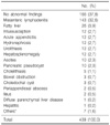
Ultrasonographic examination plays an important role in non-invasive and prompt screening examinations in detecting abdominal diseases. In this review, the author's experience of the usefulness and pitfalls of ultrasonographic examinations in children with gastrointestinal symptoms is presented. A total of 1,000 cases of children who underwent ultrasonographic evaluation in the Department of Pediatrics, Pusan National University Hospital were reviewed. The main causes leading to ultrasonographic evaluation were abdominal pain (43.9%), vomiting (17.3%), elevated liver enzymes (11.8%), and jaundice (9.8%). Abnormal ultrasonographic findings accounted for 57.9% of cases. The major abnormal findings were mesenteric lymphadenitis (29.2%), fatty liver (12.1%), hepatitis (6.4%), hepatosplenomegaly (6.2%), and acute appendicitis (4.8%). The major findings in children with abdominal pain were mesenteric lymphadenitis (32.6%), intussusception (2.7%), and acute appendicitis (2.7%). The major findings in children with vomiting were mesenteric lymphadenitis (12.7%), hypertrophic pyloric stenosis (10.4%), acute appendicitis (3.5%). The major ultrasonographic findings in children with urinary tract diseases were hydronephrosis (45.4%), urolithiasis (21.5%) and cystic renal disease (18.1%). Ultrasonography performed by pediatricians is advantageous because pediatricians are able to perform the procedure with clinical information at the right time.
Figures and Tables
References
1. Choi BI. Ultrasound diagnosis of upper abdomen. 1997. 1st ed. Seoul: Ilchokak;1–273.
2. Auh YH. Abdominal ultrasound examination. J Korean Acad Fam Med. 1992. 13:469–483.
3. Han KM, Kim JH, Bae CY, Shin DH. Usefulness of abdominal ultrasonograms in health screen. J Korean Acad Fam Med. 1994. 15:183–190.
4. Vasavada P. Ultrasound evaluation of acute abdominal emergencies in infants and children. Radiol Clin N Am. 2004. 42:445–456.

5. Kalifa G. Pediatric ultrasonography. 1986. 2nd ed. New York: Springer-Verlag Co.
6. Cheon YJ, Jang HY, Ryu JY, Eo EK, Jung KY. Emergency ultrasonography as a primary screening tool in trauma victims. J Korean Soc Traumatol. 2001. 14:62–68.
7. Mason JD. The evaluation of acute abdominal pain in children. Emerg Med Clin North Am. 1996. 14:629–643.

8. Moon HS, Woo YN. The diagnostic efficacy of abdominal ultrasonography for evaluation of children with urinary tract infection. Korean J Urol. 1996. 37:979–985.
9. Hayden CK Jr. Ultrasonography of the acute pediatric abdomen. Radiol Clin North Am. 1996. 34:791–806.
10. Park CH, Lee DH, Kim HL, Park JM, Hwang JB, Kim HS, et al. Clinical observation of mesenteric lymphadenitis in children. Korean J Pediatr. 2004. 47:31–35.
11. Sivit CJ, Newman KD, Chandra RS. Visualization of enlarged mesenteric lymph nodes at US examination. Clinical significance. Pediatr Radiol. 1993. 23:471–475.

12. Lim HT, Park JK, Choi HJ, Kim JS, Shin HK, Gu CH. Clinical approach of ultrasonography in the diagnosis of intussusception in infant and children. J Korean Pediatr Soc. 1994. 37:649–654.
13. Koumanidou C, Vakaki M, Pitsoulakis G, Kakavakis K, Mirilas P. Sonographic detection of lymph nodes in the intussusception of infant and young children: reliability of US in diagnosis. Radiology. 1994. 191:781–785.
14. Lee MK, Im CS, An SM, Kim CH, Lee DJ, Kwon JH. Ultrasonography for diagnosis of acute appendicitis in children. J Korean Pediatr Soc. 1996. 39:497–502.
15. Smith DE, Kirchmer NA, Stewart DR. Use of the barium enema in the diagnosis of acute appendicitis and its complications. Am J Surg. 1979. 138:829–834.

16. Jung TY, Choi DH, Lee CW. A clinical analysis of the appendicitis in children. J Korean Surg Soc. 1992. 43:767–775.
17. Seo HS, Chung MH, Kim KT. Ultrasonography for the acute appendicitis. J Korean Radiol Soc. 1987. 23:998–1007.

18. Lee JD, Lee JD, Cho JH, Yang JY. Diagnosis of acute appendicitis by ultrasonography. Korean Soc Ultrasound Med. 1987. 6:158–167.
19. Abu-Yousef MM, Bleicher JJ, Maber JW, Urdaneta LF, Franker FA, Metcalf AM. High-resolution sonography of acute appendicitis. Am J Radiol. 1987. 149:53–58.

20. Rubin SZ, Martin DJ. Ultrasonography in the management of possible appendicitis in childhood. J Pediatr Surg. 1990. 25:737–740.

21. Kim JH, Kim WT, Jung BO. Significance between ultrasonographic and operative findings in hypertrophic pyloric stenosis. J Korean Pediatr Soc. 2001. 44:426–432.
22. Kang SH, Lee C, Nam GR, Han DK. Ultrasonographic diagnosis of congenital hypertrophic pyloric stenosis. J Korean Pediatr Soc. 1989. 32:756–764.
23. Lee SK, Oh JW, Oh YK, Kim CG. Ultrasonographic diagnosis by pyloric volume measurement in congenital hypertrophic pyloric stenosis. J Korean Pediatr Soc. 1994. 37:1595–1599.
24. Stunden RJ, LeQuesne GW, Little KE. The improved ultrasound diagnosis of hypertrophic pyloric stenosis. Pediatr Radiol. 1986. 16:200–205.

25. Keller H, Waldmann D, Greiner P. Comparison of preoperative sonography with intraoperative findings in congenital hypertrophic pyloric stenosis. J Pediatr Surg. 1987. 22:950–952.

26. Cho KH, Hong MH, Yu HD, Lee TH, Cho AK, Park YK, et al. Clinical significance of fatty liver diagnosed by abdominal ultrasonography. J Korean Acad Fam Med. 1993. 14:734–742.
27. Kim SH, Kang DH, Lee SH, Yun CH. Causes of fatty liver diagnosed by abdominal ultrasonography. J Korean Acad Fam Med. 1995. 16:785–794.


 PDF
PDF ePub
ePub Citation
 Print
Print


 XML Download