

 PDF
PDF ePub
ePub Citation
 Print
Print


Abstract
Purpose
Visceral adipose tissue may be strongly linked to increased metabolic risks in adults. However, because little is known regarding the effect of visceral adipose tissue in children and adolescents, we performed this study to determine the association between abdominal fat distribution and metabolic risk factors in this population.
Methods
One hundred one children and adolescents (78 males and 23 females; mean age, 10.8±2.4 years) were enrolled. The anthropometric data and metabolic risk factors were evaluated. Theabdominal fat distribution was assessed according to the CT measurement. Age-adjusted, partial correlations were performed among the visceral adipose fat area (VFA), subcutaneous adiposefat area (SFA), metabolic risk factors, and anthropometrics.
Results
The SFA increased more rapidly than the VFA with advancing years in both genders. In males, the VFA and SFA were positively correlated with anthropometrics. The VFA was correlated with low HDL-cholesterol and the SFA was correlated with diastolic blood pressure (DBP). However, there was no statistical significance between the VFA, SFA, anthropometrics, and other metabolic risk factors. The VFA and SFA were strongly linked to a number of metabolic risk factors, such as other anthropometrics.
Conclusion
This study investigated how a low HDL-C was correlated with VFA and how a high DBP was associated with SFA in Korean male children and adolescents. Our results suggest that the correlation between the VFA, SFA, and metabolic risk factors was relatively weak compared to that reported in previous adult studies.
Figures and Tables
Fig. 1
Scatter plots showed the visceral fat area (VFA) and subcutaneous fat area (SFA) according to age and gender (left, male right, female). The SFA increased more rapidly compared to the VFA with advancing years in both genders.
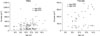
Fig. 2
Scatter plots showed a negative linear association between the visceral fat area (VFA) and HDL-cholesterol. Age-adjusted multivariate regression model identified R2 as 0.322 and p-value as <0.001 between logVFA and HDL-cholesterol.
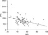
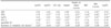
Table 1
Clinical Characteristics, Plasma Biochemistry and Radiologic Assessment of the VFA and SFA Volume of the Participants
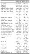
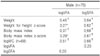
Table 3
Partial Correlations Adjusted for Age between Radiologic Assessment of the VFA, SFA, Anthropometric Data and Metabolic Risk Factors in 66 Males*
*Twelve patients were excluded lack of WC and blood pressure data, †p<0.05, ‡p<0.01, §p<0.001, ∥Metabolic risk factors include BMI above the 97th percentile (z-score,2.0 or more), a triglyceride level above the 95th percentile, an HDL cholesterol level below the 5th percentile, systolic or diastolic blood pressure above the 95th percentile, and impaired glucose tolerance, WC: waist circumference, SBP: systolic blood pressure, DBP: diastolic blood pressure, TG: triglyceride, HDL-C: high density lipoprotein cholesterol.
References
1. Ogden CL, Carroll MD, Curtin LR, McDowell MA, Tabak CJ, Flegal KM. Prevalence of overweight and obesity in the United States, 1999-2004. JAMA. 2006. 295:1549–1555.

2. Oh K, Jang MJ, Lee NY, Moon JS, Lee CG, Yoo MH, et al. Prevalence and trends in obesity among Korean children and adolescents in 1997 and 2005. Korean J Pediatr. 2008. 51:950–955.
3. Sinaiko AR, Donahue RP, Jacobs DR Jr, Prineas RJ. Relation of weight and rate of increase in weight during childhood and adolescence to body size, blood pressure, fasting insulin, and lipids in young adults. The Minneapolis Children's Blood Pressure Study. Circulation. 1999. 99:1471–1476.
4. Barton M. Screening for obesity in children and adolescents: US Preventive Services Task Force recommendation statement. Pediatrics. 2010. 125:361–367.
5. Lee S, Bacha F, Gungor N, Arslanian SA. Waist circumference is an independent predictor of insulin resistance in black and white youths. J Pediatr. 2006. 148:188–194.
6. Zimmet P, Alberti KG, Kaufman F, Tajima N, Silink M, Arslanian S, et al. The metabolic syndrome in children and adolescents - an IDF consensus report. Pediatr Diabetes. 2007. 8:299–306.
7. Park MJ, Boston BA, Oh M, Jee SH. Prevalence and trends of metabolic syndrome among Korean adolescents: from the Korean NHANES survey, 1998~2005. J Pediatr. 2009. 155:529–534.
8. Wajchenberg BL. Subcutaneous and visceral adipose tissue: their relation to the metabolic syndrome. Endocr Rev. 2000. 21:697–738.
9. Fox CS, Massaro JM, Hoffmann U, Pou KM, Maurovich-Horvat P, Liu CY, et al. Abdominal visceral and subcutaneous adipose tissue compartments: association with metabolic risk factors in the Framingham Heart Study. Circulation. 2007. 116:39–48.
10. Matsushita Y, Nakagawa T, Yamamoto S, Takahashi Y, Yokoyama T, Noda M, et al. Associations of visceral and subcutaneous fat areas with the prevalence of metabolic risk factor clustering in 6292 Japanese individuals: the Hitachi Health Study. Diabetes Care. 2010. 05. 11. [Epub ahead of print]. doi: 10.2337/dc10-0120.
11. Moon JS, Lee SY, Nam CM, Choi JM, Choe BK, Seo JW, et al. 2007 Korean National Growth Charts: review of developmental process and an outlook. Korean J Pediatr. 2008. 51:1–25.
12. Cole TJ, Green PJ. Smoothing reference centile curves: the LMS method and penalized likelihood. Stat Med. 1992. 11:1305–1319.
13. Matthews DR, Hosker JP, Rudenski AS, Naylor BA, Treacher DF, Turner RC. Homeostasis model assessment: insulin resistance and beta-cell function from fasting plasma glucose and insulin concentrations in man. Diabetologia. 1985. 28:412–419.
14. Weiss R, Dziura J, Burgert TS, Tamborlane WV, Taksali SE, Yeckel CW, et al. Obesity and the metabolic syndrome in children and adolescents. N Engl J Med. 2004. 350:2362–2374.
15. Bjorntorp P. "Portal" adipose tissue as a generator of risk factors for cardiovascular disease and diabetes. Arteriosclerosis. 1990. 10:493–496.
16. Fontana L, Eagon JC, Trujillo ME, Scherer PE, Klein S. Visceral fat adipokine secretion is associated with systemic inflammation in obese humans. Diabetes. 2007. 56:1010–1013.
17. Caprio S, Hyman LD, McCarthy S, Lange R, Bronson M, Tamborlane WV. Fat distribution and cardiovascular risk factors in obese adolescent girls: importance of the intraabdominal fat depot. Am J Clin Nutr. 1996. 64:12–17.
18. Suliga E. Visceral adipose tissue in children and adolescents: a review. Nutr Res Rev. 2009. 22:137–147.
19. Kim JA, Park HS. Association of abdominal fat distribution and cardiometabolic risk factors among obese Korean adolescents. Diabetes Metab. 2008. 34:126–130.
20. Kahn SE, Hull RL, Utzschneider KM. Mechanisms linking obesity to insulin resistance and type 2 diabetes. Nature. 2006. 444:840–846.
21. Fox KR, Peters DM, Sharpe P, Bell M. Assessment of abdominal fat development in young adolescents using magnetic resonance imaging. Int J Obes Relat Metab Disord. 2000. 24:1653–1659.
22. Kim JA, Ju SY, Yum KS. Cut-off value of visceral fat area at risk of obesity-related disorders in Korean adult population. J Korean Acad Fam Med. 2006. 27:208–214.
23. Jackson AS, Pollock ML, Graves JE, Mahar MT. Reliability and validity of bioelectrical impedance in determining body composition. J Appl Physiol. 1988. 64:529–534.
24. Kullberg J, Brandberg J, Angelhed JE, Frimmel H, Bergelin E, Strid L, et al. Whole-body adipose tissue analysis: comparison of MRI, CT and dual energy X-ray absorptiometry. Br J Radiol. 2009. 82:123–130.
25. Stolk RP, Wink O, Zelissen PM, Meijer R, van Gils AP, Grobbee DE. Validity and reproducibility of ultrasonography for the measurement of intra-abdominal adipose tissue. Int J Obes Relat Metab Disord. 2001. 25:1346–1351.
26. Blouin K, Boivin A, Tchernof A. Androgens and body fat distribution. J Steroid Biochem Mol Biol. 2008. 108:272–280.
 XML Download