

 PDF
PDF ePub
ePub Citation
 Print
Print


Abstract
Fractures and fracture-dislocations of the ankle are caused by a variety of mechanisms. In addition to fractures, injuries of soft tissue, such as ligaments, tendons, nerves, and muscles may also occur. Among these, a tibialis posterior tendon injury is difficult to be identified due to swelling and pain at the fracture site. It is difficult to observe tibialis posterior tendon injury on a simple radiograph; it is usually found during surgery by accident. There are some studies regarding irreducible ankle fracture-dislocations due to interposition of the tibialis posterior tendon; however, to the best of our knowledge, there has not been any report about interposition of injured tibialis posterior tendon. Herein, we report a case of an irreducible fracture-dislocation of the ankle due to injured tibialis posterior tendon interposition that was observed intraoperatively, interrupting the reduction of ankle fracture-dislocation. We obtained satisfactory clinical result after reduction of the trapped tendon, fracture reduction, and internal fixation; therefore, we are willing to report this case with the consent of the patient. This study was conducted with an approval from the local Institutional Ethics Review Board.
REFERENCES
1.Ermis MN., Yagmurlu MF., Kilinc AS., Karakas ES. Irreducible fracture dislocation of the ankle caused by tibialis posterior tendon interposition. J Foot Ankle Surg. 2010. 49:111–71.

2.Lacasse JS., Laflamme M., Penner MJ. Irreducible fracture-dislocation of the ankle associated with interposition of the tibialis posterior tendon in the syndesmosis: a case report. J Foot Ankle Surg. 2015. 54:912–1.
3.Jarvis HC., Cannada LK. Acute tibialis posterior tendon rupture associated with a distal tibial fracture. Orthopedics. 2012. 35:e595–7.
4.Walker RH., Farris C. Irreducible fracture-dislocations of the ankle associated with interposition of the tibialis posterior tendon: case report and review of the literature of a specific ankle fracture syndrome. Clin Orthop Relat Res. 1981. 110:212–1.
5.Hunter AM., Bowlin C. Posterior tibial tendon entrapment within an intact ankle mortise: a case report. J Foot Ankle Surg. 2015. 54:111–9.
6.Giblin MM. Ruptured tibialis posterior tendon associated with a closed medial malleolar fracture. Aust N Z J Surg. 1980. 50:59–10.
7.West MA., Sangani C., Toh E. Tibialis posterior tendon rupture associated with a closed medial malleolar fracture: a case report and review of the literature. J Foot Ankle Surg. 2010. 49:515. .e9-12.
8.Coonrad RW., Bugg EI Jr. Trapping of the posterior tibial tendon and interposition of soft tissue in severe fractures about the ankle joint. J Bone Joint Surg Am. 1954. 31:744–50.
9.Cunningham DJ., Romanes GJ. Cunningham’s textbook of anatomy. 12th ed.London: Oxford University Press;1981.
10.Holmes GB Jr., Mann RA. Possible epidemiological factors associated with rupture of the posterior tibial tendon. Foot Ankle. 1992. 13:70–9.
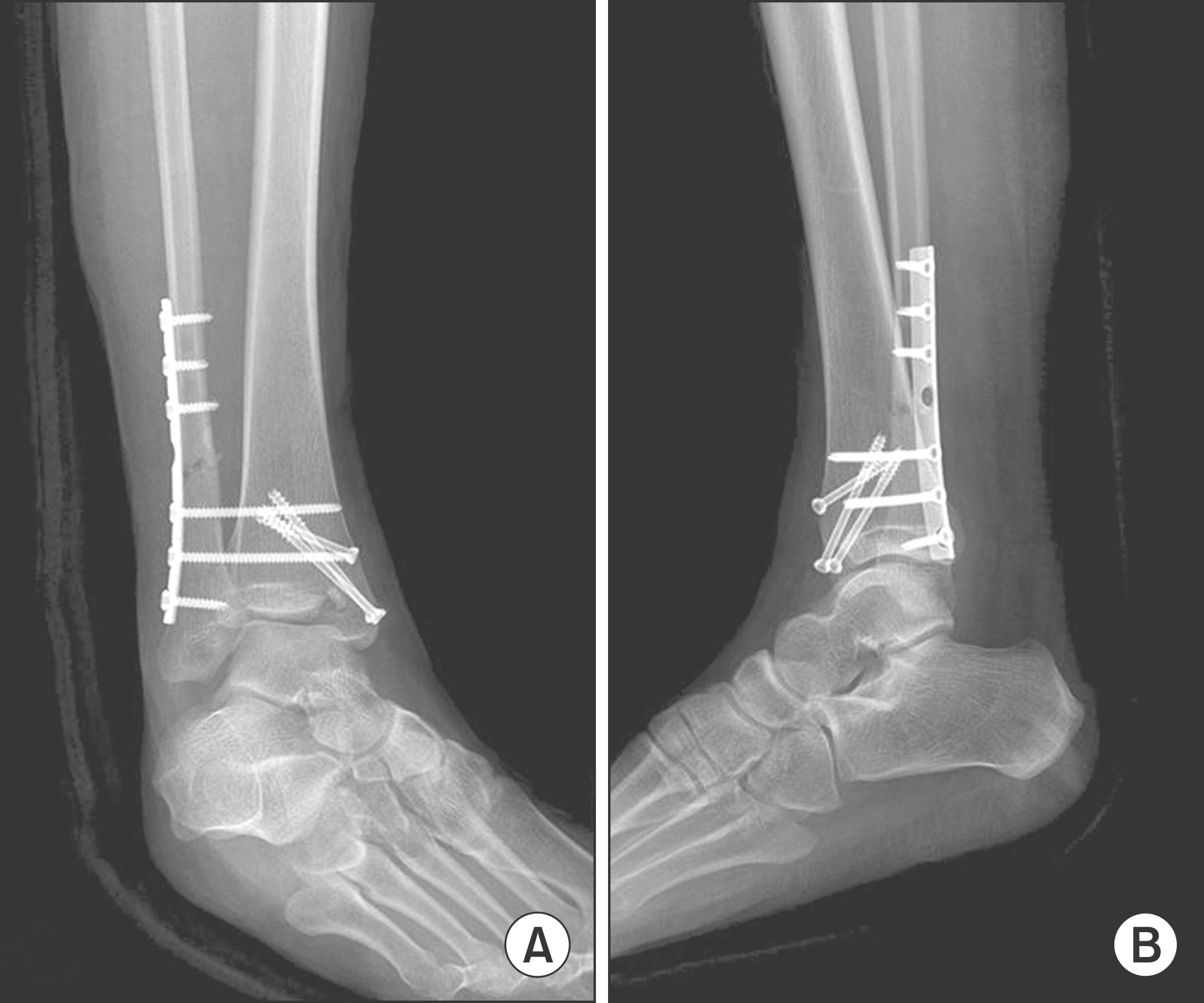
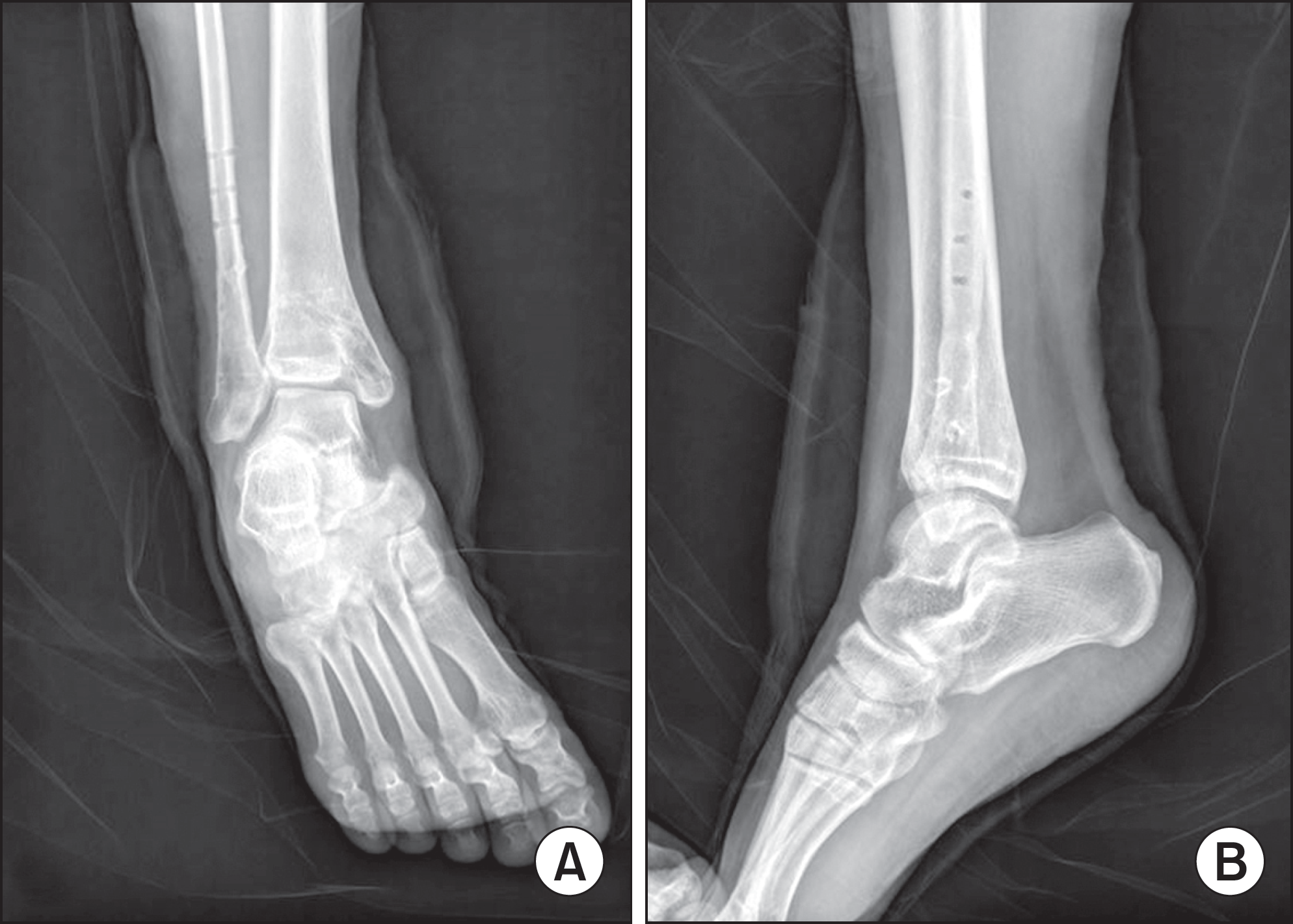
Figure 1.
Preoperative radiographs showed AO-OTA (Arbeitsgemein-schaft für osteosynthesefragen-Orthopedic Trauma Association) type 44-B2, ankle fracture. Lateral subluxation of talus and widening of syndemosis were observed. (A) Mortise view. (B) Lateral view.
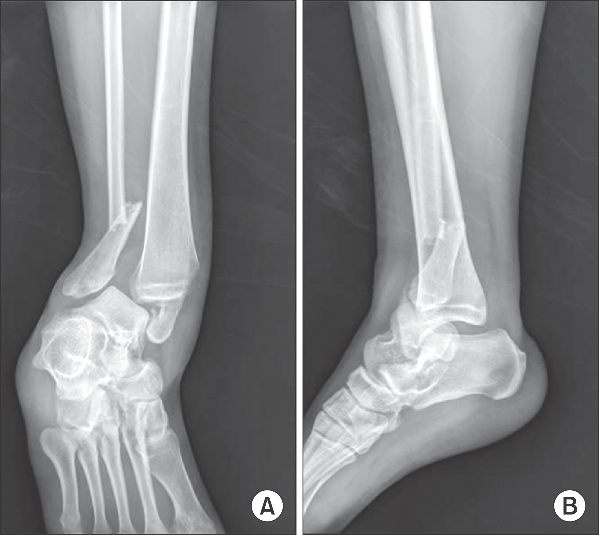
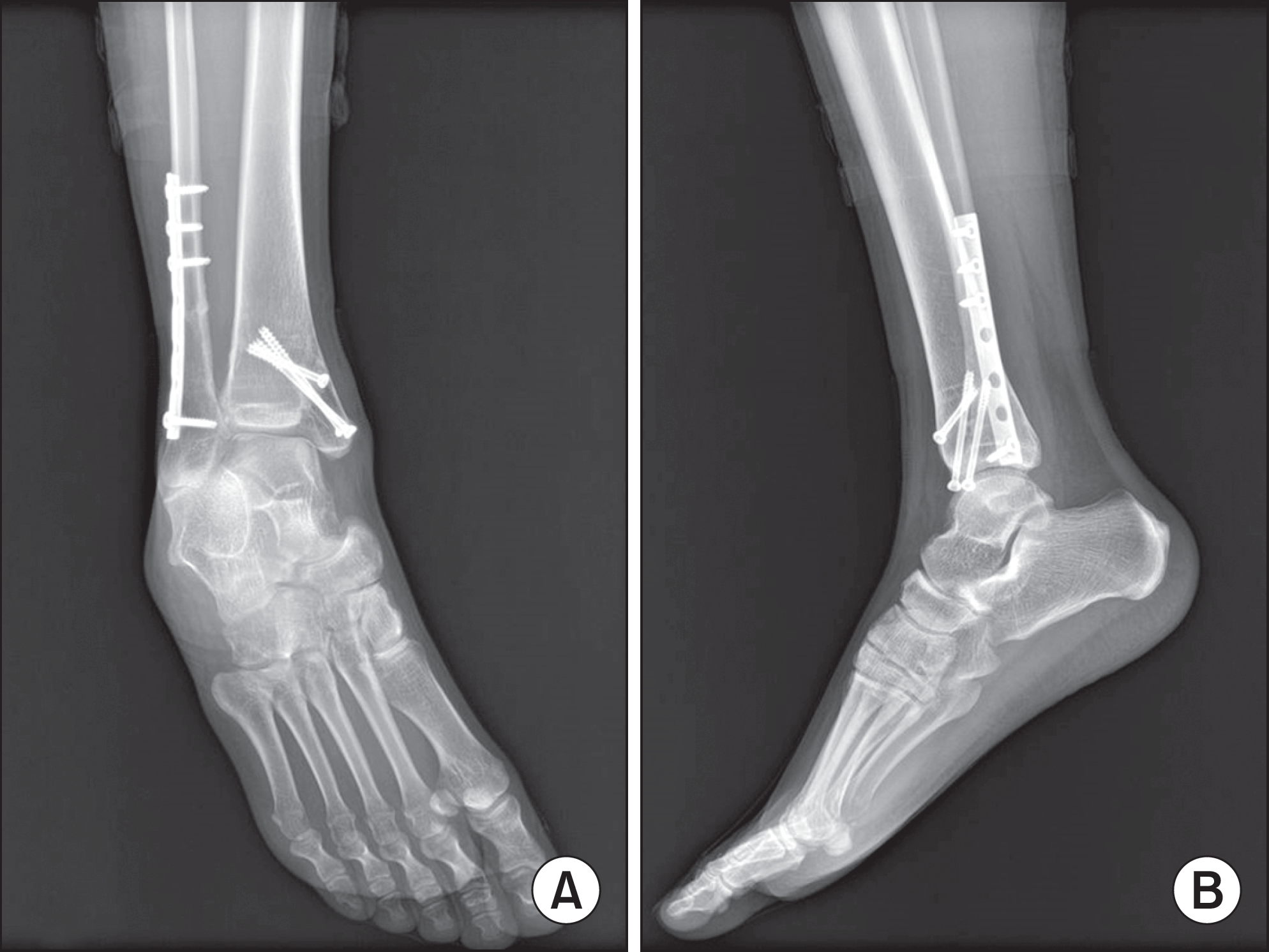
Figure 2.
Irreducible ankle fracture and dislocation after manual reduction. (A) Mortise view. (B) Lateral view.
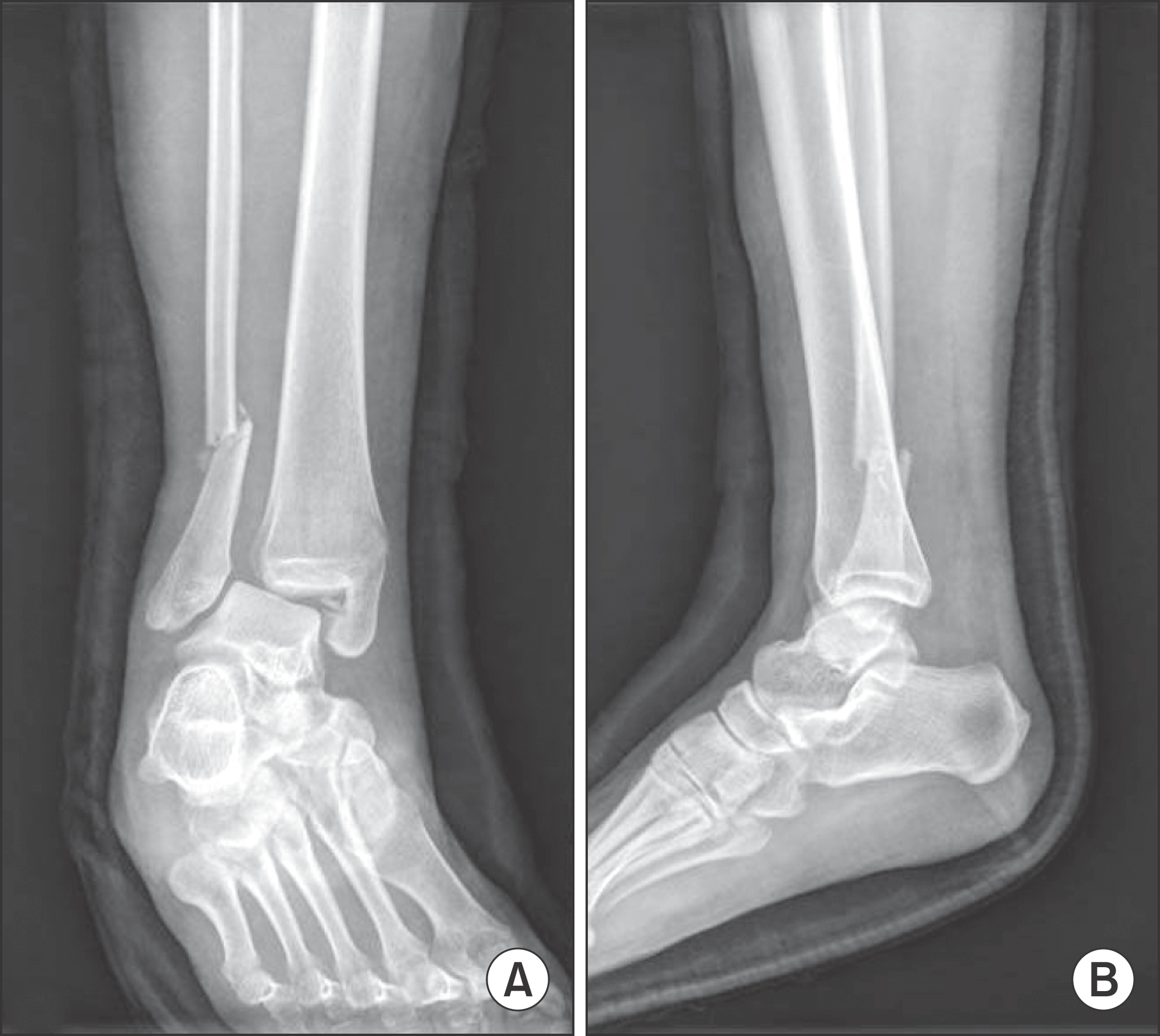
Figure 3.
Intraoperative fluoroscopic image showed that the reduction was satisfactory and ankle joint congruency was well maintained.
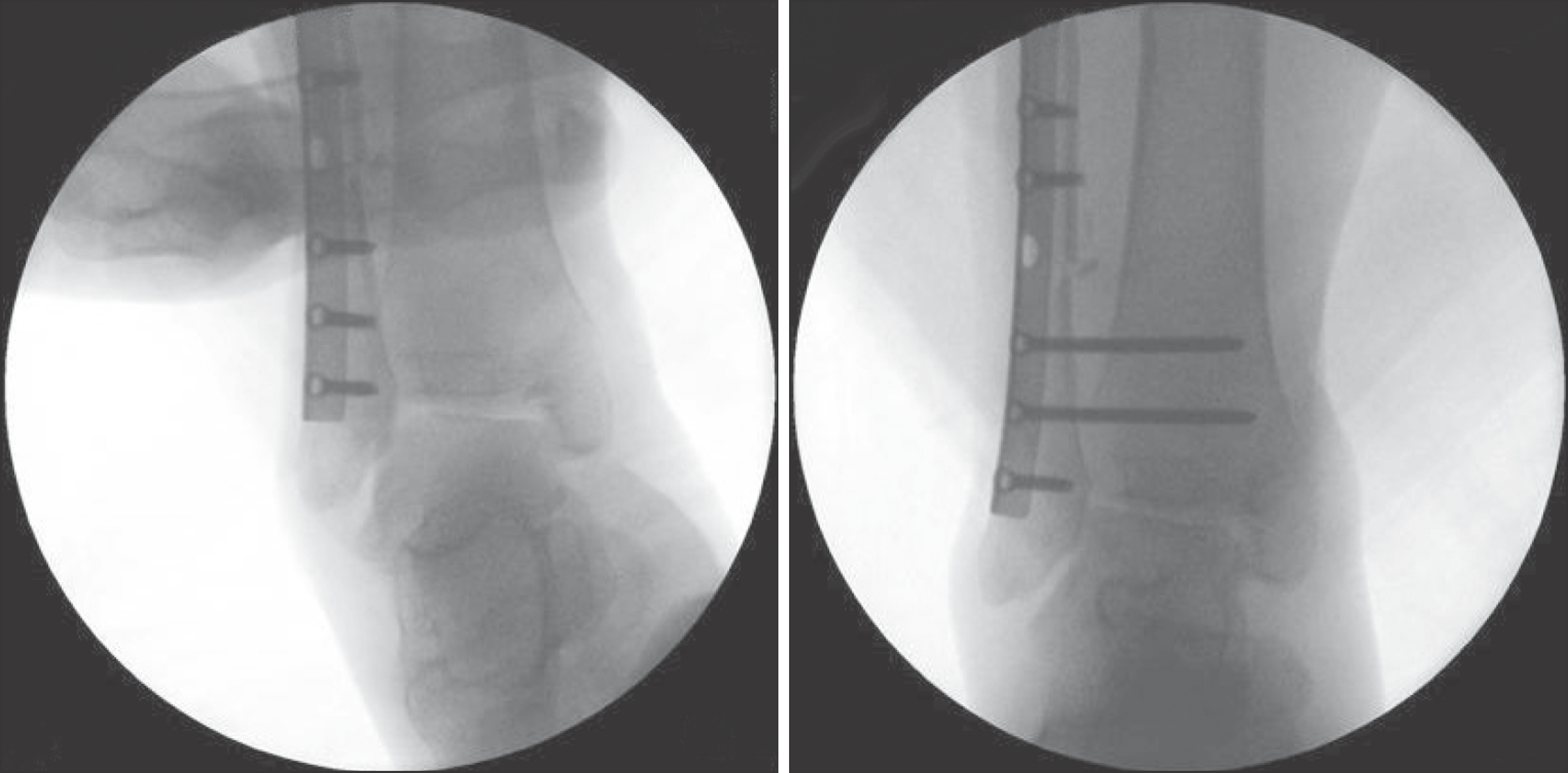
Figure 4.
Intraoperative fluoroscopic image showed that the reduction of medial malleolar fracture was not satisfactory and ankle mortise was not maintained.
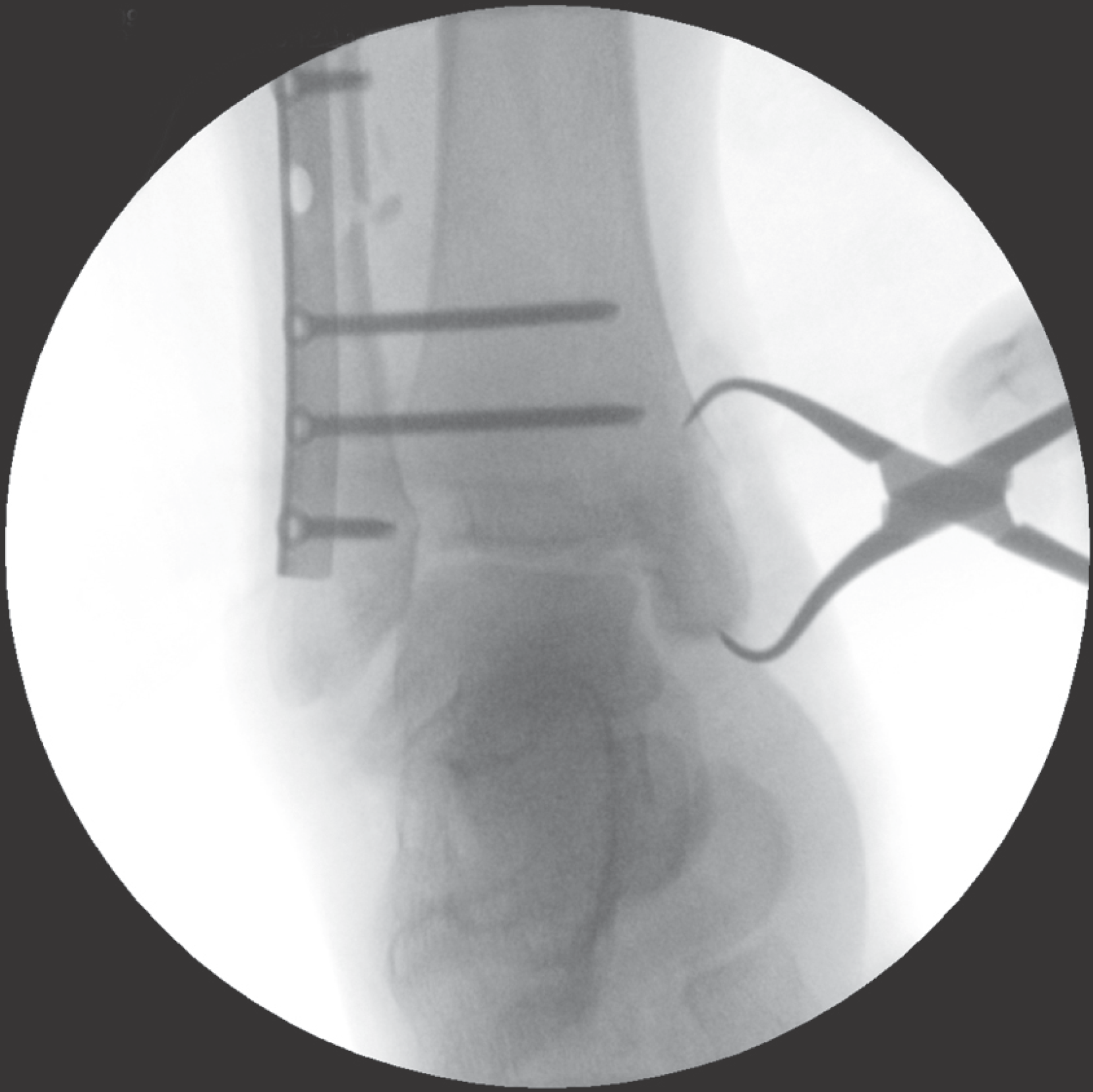
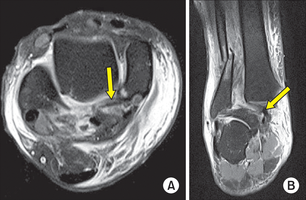
Figure 6.
Three-dimensional reconstruction images of operative findings. Tibialis posterior tendon (arrows) was passing by posterior surface of talus between the distal tibia and fibula and from lateral to medial.

Figure 5.
Ankle joint findings during operation. (A) Tibialis posterior tendon was found at intra-articular space of ankle. (B) Anatomical reduction was inhibited by interposition of tibialis posterior tendon. (C) Partially ruptured tibialis posterior tendon was observed.
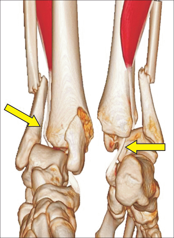
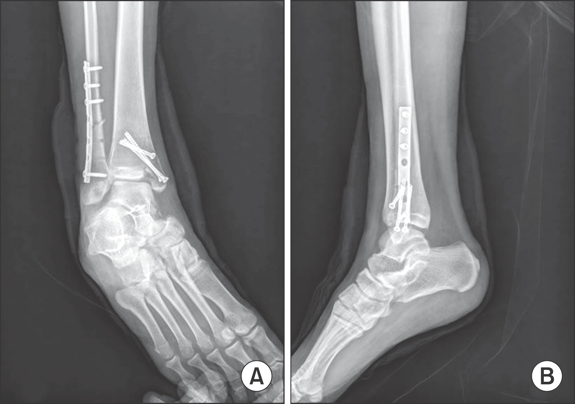
Figure 7.
Postoperative radiographs after operation showed well, satisfactory reduction. (A) Mortise view. (B) Lateral view.
 XML Download