

 PDF
PDF ePub
ePub Citation
 Print
Print


Abstract
Purpose:
Materials and Methods:
Results:
REFERENCES
Figure 1.
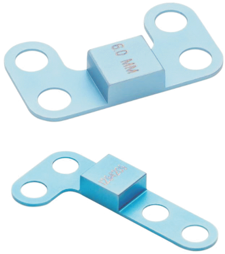
Figure 2.

Figure 3.

Table 1.
| Variable | Group C (n=12) | Group MT (n=12) |
|---|---|---|
| Mean age (yr) | 37.1±13.18 | 43.14±14.25 |
| Follow-up period (mo) | 16.22±4.16 | 17.13±4.27 |
| Male:female | 4:8 | 3:9 |
| Right:left | 4:8 | 8:4 |
Table 2.
Values are presented as mean±standard deviation. Group C: Cotton osteotomy, Group MT: 1st metatarsal base osteotomy, Preop: preoperative, Postop: postoperative, TNCA: talo-navicular coverage angle on anteroposterior (AP) radiograph, T1MTA: talo-1st metatarsal angle on AP radiograph, TCA: talo-calcaneal angle on lateral radiograph, CPA: calcaneal pitch angle on lateral radiograph, MCH: medial cuneiform height on lateral radiograph, AOFAS: American Orthopaedic Foot and Ankle Society.
Table 3.
Values are presented as mean±standard deviation. Group C: Cotton osteotomy, Group MT: 1st metatarsal base osteotomy, Δ: delta, an increment of a variable, TNCA: talo-navicular coverage angle on anteroposterior (AP) radiograph, T1MTA: talo-1st metatarsal angle on AP radiograph, TCA: talo-calcaneal angle on lateral radiograph, CPA: calcaneal pitch angle on lateral radiograph, MCH: medial cuneiform height on lateral radiograph.
 XML Download