

 PDF
PDF ePub
ePub Citation
 Print
Print


Abstract
Hallux valgus is a deformity characterized by lateral deviation of the great toe and medial deviation of the first metatarsal. When planning an operative treatment, it is important to realize that the deformity is tridimensional and diverse. Operative techniques include medial eminence resection, distal soft tissue procedure, first metatarsal osteotomy (distal, diaphyseal, proximal, or multiple), proximal phalanx osteotomy, arthrodesis (first metatarsophalangeal or metatarsocuneiform joint), and so on. Among these techniques, osteotomy is the main procedure for correcting the hallux valgus. The objective of this article is to describe the characteristics and recent advancements made for corrective osteotomies in the hallux valgus. The pathophysiology of the hallux valgus is also described.
Figures and Tables
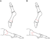
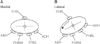 | Figure 1Schematic representation of tendons around the first metatarsal head. (A) Normal articulation in a balanced state. (B) Relationship of the tendons in hallux valgus deformity. EHB: extensor hallucis brevis, ABH: abductor hallucis, FHBM: medial head of flexor hallucis brevis, FHBL: lateral head of flexor hallucis brevis, ADH: adductor hallucis.
|
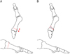
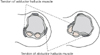 | Figure 2Relationship of the sesamoids to the metatarsal head. Left: the sesamoids stabilized by the crista. Right: atrophy of the crista with lateral deviation of the sesamoids.
|
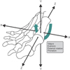

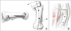 | Figure 7(A, B) The modified rotational scarf procedure rotates the two halves of the osteotomy, assuring adequate cortical crossover and thereby eliminates linear overlap between the cortices of the first metatarsal and softer cancellous bone (used with permission from TriMed Inc., Santa Clarita, CA, USA).
|
References
1. Coughlin MJ, Jones CP. Hallux valgus: demographics, etiology, and radiographic assessment. Foot Ankle Int. 2007; 28:759–777.

2. Mann RA, Rudicel S, Graves SC. Repair of hallux valgus with a distal soft-tissue procedure and proximal metatarsal osteotomy. A long-term follow-up. J Bone Joint Surg Am. 1992; 74:124–129.
3. Scranton PE Jr, Rutkowski R. Anatomic variations in the first ray: Part I. Anatomic aspects related to bunion surgery. Clin Orthop Relat Res. 1980; (151):244–255.
4. Dayton P, Kauwe M, Feilmeier M. Clarification of the anatomic definition of the bunion deformity. J Foot Ankle Surg. 2014; 53:160–163.
5. Glynn MK, Dunlop JB, Fitzpatrick D. The Mitchell distal metatarsal osteotomy for hallux valgus. J Bone Joint Surg Br. 1980; 62:188–191.
6. Hardy RH, Clapham JC. Observations on hallux valgus; based on a controlled series. J Bone Joint Surg Br. 1951; 33:376–391.
7. Coughlin MJ. Juvenile hallux valgus: etiology and treatment. Foot Ankle Int. 1995; 16:682–697.
8. Sim-Fook L, Hodgson AR. A comparison of foot forms among the non-shoe and shoe-wearing Chinese population. J Bone Joint Surg Am. 1958; 40:1058–1062.
9. Kato T, Watanabe S. The etiology of hallux valgus in Japan. Clin Orthop Relat Res. 1981; (157):78–81.
10. Munteanu SE, Menz HB, Wark JD, Christie JJ, Scurrah KJ, Bui M, et al. Hallux valgus, by nature or nurture? A twin study. Arthritis Care Res (Hoboken). 2016; 11. 18. DOI: 10.1002/acr.23154.
11. Coughlin MJ. Hallux valgus. J Bone Joint Surg Am. 1996; 78:932–966.
12. Willegger M, Holinka J, Ristl R, Wanivenhaus AH, Windhager R, Schuh R. Correction power and complications of first tarsometatarsal joint arthrodesis for hallux valgus deformity. Int Orthop. 2015; 39:467–476.
13. Schuh R, Willegger M, Holinka J, Ristl R, Windhager R, Wanivenhaus AH. Angular correction and complications of proximal first metatarsal osteotomies for hallux valgus deformity. Int Orthop. 2013; 37:1771–1780.
14. Smith SE, Landorf KB, Butterworth PA, Menz HB. Scarf versus chevron osteotomy for the correction of 1-2 intermetatarsal angle in hallux valgus: a systematic review and meta-analysis. J Foot Ankle Surg. 2012; 51:437–444.
15. Faour-Martín O, Martín-Ferrero MA, Valverde García JA, Vega-Castrillo A, de la. Long-term results of the retrocapital metatarsal percutaneous osteotomy for hallux valgus. Int Orthop. 2013; 37:1799–1803.
16. Lucas y Hernandez J, Golanó P, Roshan-Zamir S, Darcel V, Chauveaux D, Laffenêtre O. Treatment of moderate hallux valgus by percutaneous, extra-articular reverse-L Chevron (PERC) osteotomy. Bone Joint J. 2016; 98:365–373.
17. Díaz Fernández R. Percutaneous triple and double osteotomies for the treatment of hallux valgus. Foot Ankle Int. 2017; 38:159–166.
18. Badwey TM, Dutkowsky JP, Graves SC, Richardson EG. An anatomical basis for the degree of displacement of the distal chevron osteotomy in the treatment of hallux valgus. Foot Ankle Int. 1997; 18:213–215.
19. Park CH, Jang JH, Lee SH, Lee WC. A comparison of proximal and distal chevron osteotomy for the correction of moderate hallux valgus deformity. Bone Joint J. 2013; 95:649–656.
20. Park HW, Lee KB, Chung JY, Kim MS. Comparison of outcomes between proximal and distal chevron osteotomy, both with supplementary lateral soft-tissue release, for severe hallux valgus deformity: a prospective randomised controlled trial. Bone Joint J. 2013; 95:510–516.
21. Bai LB, Lee KB, Seo CY, Song EK, Yoon TR. Distal chevron osteotomy with distal soft tissue procedure for moderate to severe hallux valgus deformity. Foot Ankle Int. 2010; 31:683–688.
22. Resch S, Stenstrom A, Gustafson T. Circulatory disturbance of the first metatarsal head after Chevron osteotomy as shown by bone scintigraphy. Foot Ankle. 1992; 13:137–142.
23. Garrido IM, Rubio ER, Bosch MN, González MS, Paz GB, Llabrés AJ. Scarf and Akin osteotomies for moderate and severe hallux valgus: clinical and radiographic results. Foot Ankle Surg. 2008; 14:194–203.
24. Adam SP, Choung SC, Gu Y, O'Malley MJ. Outcomes after scarf osteotomy for treatment of adult hallux valgus deformity. Clin Orthop Relat Res. 2011; 469:854–859.
25. Aminian A, Kelikian A, Moen T. Scarf osteotomy for hallux valgus deformity: an intermediate followup of clinical and radiographic outcomes. Foot Ankle Int. 2006; 27:883–886.
26. Coetzee JC, Rippstein P. Surgical strategies: scarf osteotomy for hallux valgus. Foot Ankle Int. 2007; 28:529–535.
27. Murawski CD, Egan CJ, Kennedy JG. A rotational scarf osteotomy decreases troughing when treating hallux valgus. Clin Orthop Relat Res. 2011; 469:847–853.
28. Seng C, Chunyin Ho D, Chong KW. Restoring sesamoid position in scarf osteotomy: a learning curve. J Foot Ankle Surg. 2015; 54:1089–1092.
29. Castaneda DA, Myerson MS, Neufeld SK. The Ludloff osteotomy: a review of current concepts. Int Orthop. 2013; 37:1661–1668.
30. Saxena A, St Louis M. Medial locking plate versus screw fixation for fixation of the Ludloff osteotomy. J Foot Ankle Surg. 2013; 52:153–157.
31. Beischer AD, Ammon P, Corniou A, Myerson M. Three-dimensional computer analysis of the modified Ludloff osteotomy. Foot Ankle Int. 2005; 26:627–632.
32. Iyer S, Demetracopoulos CA, Sofka CM, Ellis SJ. High rate of recurrence following proximal medial opening wedge osteotomy for correction of moderate hallux valgus. Foot Ankle Int. 2015; 36:756–763.
33. Yasuda T, Okuda R, Jotoku T, Shima H, Hida T, Neo M. Proximal supination osteotomy of the first metatarsal for hallux valgus. Foot Ankle Int. 2015; 36:696–704.
34. Goldbloom D, Makwana N, Laing P, Toullec E, Graff W, Charbel A. A new “tension side”" locking plate for Hallux Valgus: a prospective multicentre case series. Foot Ankle Surg. 2016; 22:103–108.
35. Nyska M. Principles of first metatarsal osteotomies. Foot Ankle Clin. 2001; 6:399–408.
36. Smith BW, Coughlin MJ. Treatment of hallux valgus with increased distal metatarsal articular angle: use of double and triple osteotomies. Foot Ankle Clin. 2009; 14:369–382.
37. Jeyaseelan L, Chandrashekar S, Mulligan A, Bosman HA, Watson AJ. Correction of moderate to severe hallux valgus with combined proximal opening wedge and distal chevron osteotomies: a reliable technique. Bone Joint J. 2016; 98:1202–1207.
38. Tóth K, Kellermann P, Wellinger K. Fixation of Akin osteotomy for hallux abductus with absorbable suture. Arch Orthop Trauma Surg. 2010; 130:1257–1261.
39. Sinnett T, Fang Y, Nattfogel E, O'Gorman A, Charalambides C. Suture fixation of an Akin osteotomy: a cost effective and clinically reliable technique. Foot Ankle Surg. 2017; 23:40–43.
 XML Download