

 PDF
PDF ePub
ePub Citation
 Print
Print


Abstract
Surgical treatments for arthritis in the first metatarsophalangeal joint include arthrodesis, interposition arthroplasty using silicone or meniscus cartilage, and rarely arthroplasty. Although arthrodesis was performed successfully, pain can persist if the angle of fusion was inappropriate. Interposition arthroplasty can be tried for the treatment of persisting pain after the arthrodesis. Interposition arthroplasty using tensor fascia lata is known that has low risk of adhesions and easy to harvest. Compared to autologous grafts, grafting rates is high and low risk of rejection additionally. Herein, we report a successfully managed arthritis with severe pain with interposition arthroplasty using tensor fascia lata after a failed metatarsophalangeal joint arthrodesis.
Figures and Tables
Figure 1
(A) Anteroposterior view of preoperative plain radiograph with standing demonstrates metatarsophalangeal angle (MPA) was 23°. (B) Lateral view of preoperative plain radiograph with standing demonstrates MPA was 15°. (C, D) Axial view of preoperative computed tomography (CT), sagittal view of preoperative CT shows union of first metatarsophalangeal joint.
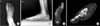
Figure 2
(A, B) Osteotomy was made from distal part of first metatarsophalangeal joint and proximal part of first metatarsophalangeal joint in dorsal side. (C) Operative finding of successful osteotomy of first metatarsophalangeal joint.
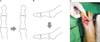
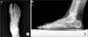
Figure 3
(A) Tensor fascia lata was harvested from lateral side of ipsilateral thigh. (B) The harvested tensor fascia lata. (C) The harvested tensor fascia lata folded in triplicate.


References
1. Hamilton WG, O'Malley MJ, Thompson FM, Kovatis PE. Roger Mann Award 1995. Capsular interposition arthroplasty for severe hallux rigidus. Foot Ankle Int. 1997; 18:68–70.
2. Hamilton GA, Ford LA, Patel S. First metatarsophalangeal joint arthrodesis and revision arthrodesis. Clin Podiatr Med Surg. 2009; 26:459–473.

3. Hahn MP, Gerhardt N, Thordarson DB. Medial capsular interpositional arthroplasty for severe hallux rigidus. Foot Ankle Int. 2009; 30:494–499.
4. Miller D, Maffulli N. Free gracilis interposition arthroplasty for severe hallux rigidus. Bull Hosp Jt Dis. 2005; 62:121–124.
5. Keiserman LS, Sammarco VJ, Sammarco GJ. Surgical treatment of the hallux rigidus. Foot Ankle Clin. 2005; 10:75–96.
6. Kennedy JG, Chow FY, Dines J, Gardner M, Bohne WH. Outcomes after interposition arthroplasty for treatment of hallux rigidus. Clin Orthop Relat Res. 2006; 445:210–215.
7. Cook E, Cook J, Rosenblum B, Landsman A, Giurini J, Basile P. Meta-analysis of first metatarsophalangeal joint implant arthroplasty. J Foot Ankle Surg. 2009; 48:180–190.
8. Lawrence BR, Thuen E. A retrospective review of the primus first MTP joint double-stemmed silicone implant. Foot Ankle Spec. 2013; 6:94–100.
9. Coughlin MJ, Shurnas PJ. Soft-tissue arthroplasty for hallux rigidus. Foot Ankle Int. 2003; 24:661–672.
10. Taghinia AH, Al-Sheikh AA, Upton J. Suture anchor suspension and fascia lata interposition arthroplasty for Basal joint arthritis of the thumb. Plast Reconstr Surg. 2008; 122:497–504.
 XML Download