

 PDF
PDF ePub
ePub Citation
 Print
Print


Abstract
Traumatic neuromas are rare benign tumors that are common after trauma or surgery and are usually accompanied by obvious symptoms of pain. Most reports show neuromas in the face, neck, and limbs, and the traumatic neuroma of the medial plantar nerve has rarely been reported. We encountered a traumatic neuroma of the medial plantar nerve after a deep laceration mimicking a foreign body granuloma. A small mass lesion was found around plantar aponeurosis with heterogeneous high signal intensity in the T2 fat suppression view and slightly enhanced intensity in the magnetic resonance imaging that suggested a foreign body granuloma. The lesion was diagnosed pathologically as a traumatic neuroma. A satisfactory clinical result was obtained after excision of the traumatic neuroma and burial of the proximal and distal stumps to the adjacent muscle at the secondary operation.
Figures and Tables
Figure 1
Axial T2-weighted fat suppression magnetic resonance imaging finding of the mass lesion. Ill-defined heterogeneous two high signal intensity lesion (asterisks) located lateral to flexor hallucis longus tendon.
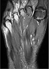
Figure 2
Ultrasonographic finding of the mass lesion. Ill-defined heterogeneous hypoechoic lesion (asterisk) located between plantar aponeurosis (PA) and flexor hallucis longus (FHL) tendon.
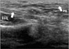
Figure 3
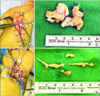
Gross photo of specimens of first surgery: two separately round-in-shape masses about 7.0×5.0 mm and 5.0×3.0 mm, respectively.
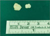
Figure 4
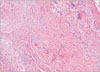
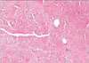
Microscopic finding of the mass lesion of first surgery. It is characterized by a variably well-defined, but unencapsulated, mass of numerous axons and Schwann cells embedded in scar tissue adjacent (H&E stain, ×100).
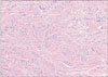
Figure 5
Operative field findings during the second operation. (A) The medial plantar nerve was (arrow) surrounded by thick fibrous tissue (asterisk). (B) After excision of the fibrous tissue, multiple fusiform masses (arrows) were founded in the medial plantar nerve trunk. (C) Gross appearance of excised fibrous tissue surrounding multiple neuromas. (D) Gross appearance of excised medial plantar nerve including multiple traumatic neuromas.
References
1. Brogan DM, Kakar S. Management of neuromas of the upper extremity. Hand Clin. 2013; 29:409–420.

2. Roh YT, Park IJ. Treatment of the traumatic neuroma. J Korean Soc Surg Hand. 2014; 19:209–220.
3. Kim J, Dellon AL. Reconstruction of a painful post-traumatic medial plantar neuroma with a bioabsorbable nerve conduit: a case report. J Foot Ankle Surg. 2001; 40:318–323.
4. Lundborg G. A 25-year perspective of peripheral nerve surgery: evolving neuroscientific concepts and clinical significance. J Hand Surg Am. 2000; 25:391–414.
5. Watson J, Gonzalez M, Romero A, Kerns J. Neuromas of the hand and upper extremity. J Hand Surg Am. 2010; 35:499–510.
6. Kang J, Yang P, Zang Q, He X. Traumatic neuroma of the superficial peroneal nerve in a patient: a case report and review of the literature. World J Surg Oncol. 2016; 14:242.
7. Wall PD, Gutnick M. Ongoing activity in peripheral nerves: the physiology and pharmacology of impulses originating from a neuroma. Exp Neurol. 1974; 43:580–593.
8. Guse DM, Moran SL. Outcomes of the surgical treatment of peripheral neuromas of the hand and forearm: a 25-year comparative outcome study. Ann Plast Surg. 2013; 71:654–658.
 XML Download