

 PDF
PDF ePub
ePub Citation
 Print
Print


Abstract
Soft tissue tumors of the foot have a low incidence rate, and most of them are symptom free, thus it is difficult to diagnose accurately. Herein, we report a 15-year-old male patient who had swelling without pain on the lateral margin of both feet. We performed excisional biopsy of the abductor digiti minimi via subtotal resection, following radiograph and magnetic resonance imaging. According to the histological analysis, hypertrophy of abductor digiti minimi was positive, and other soft tissue tumors were negative. Six months after the operation, normal appearance of both feet was maintained and the patient was satisfied with the result.
Figures and Tables
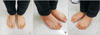
Figure 1
Soft tissue mass at the lateral aspect of the both foot was shown at first presentation. (A) Anterior view. (B) Right angular view. (C) Left angular view.
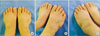
Figure 2
Diameters of the abductor digiti minimi muscles of both feet at first presentation were 20.7 mm on the left side (A) and 18.4 mm on the right side (B) on ultrasound scan.
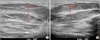
Figure 3
Magnetic resonance imaging of both feet at first presentation showed diffuse muscular swelling and clear accentuation of the abductor digiti minimi muscle in the both foot. (A) Left side. (B) Right side.
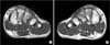
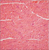
Figure 4
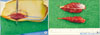
(A, B) The left side hypertrophic abductor digiti minimi muscle and the excised specimens were shown intraoperatively.
References
1. Almansoor J, Boothroyd AE, Carty H. Asymmetrical muscle hypertrophy simulating a localised soft tissue mass. J Pediatr Orthop B. 1998; 7:86–88.

2. Raab P, Ettl V, Kozuch A, Nöth U. Hypertrophy of the abductor digiti minimi muscle simulating a localised soft tissue mass. Foot Ankle Surg. 2008; 14:43–46.
3. Iconomou T, Tsoutsos D, Spyropoulou G, Gravvanis A, Ioannovich J. Congenital hypertrophy of the abductor digiti minimi muscle of the foot. Plast Reconstr Surg. 2005; 115:1223–1225.
4. Guy JA, Micheli LJ. Strength training for children and adolescents. J Am Acad Orthop Surg. 2001; 9:29–36.
5. Boland BJ, Silbert PL, Groover RV, Wollan PC, Silverstein MD. Skeletal, cardiac, and smooth muscle failure in Duchenne muscular dystrophy. Pediatr Neurol. 1996; 14:7–12.
6. Kagel KO, Ott J, Lorenz G, Mieler W. Clinical picture, symptomatology and therapy of partial angiodysplastic gigantism resembling the Klippel-Trenaunay-P.-Weber syndrome type. Zentralbl Chir. 1972; 97:50–57.
7. Boothroyd AE, Carty H. The painless soft tissue mass in childhood--tumour or not? Postgrad Med J. 1995; 71:10–16.
8. Montgomery F, Miller R. Hypertrophic extensor digitorum brevis muscles simulating pseudotumors: a case report. Foot Ankle Int. 1998; 19:566–567.
9. Ringelman PR, Goldberg NH. Hypertrophy of the abductor hallicus muscle: an unusual congenital foot mass. Foot Ankle. 1993; 14:366–369.
10. Dunn AW. Anomalous muscles simulating soft-tissue tumors in the lower extremities. Report of three cases. J Bone Joint Surg Am. 1965; 47:1397–1400.
 XML Download