

 PDF
PDF ePub
ePub Citation
 Print
Print


Abstract
Purpose:
Anterior drawer and varus stress radiographs are commonly to diagnose chronic lateral ankle instability. We compared the preoperative stress radiographs with the intraoperative radiographs under anesthesia to determine the accuracy and efficacy of stress radiographs in an outpatient clinical environment.
Materials and Methods:
Data was collected from patients who underwent a modified Broström operation for painful chronic unilateral lateral ankle instability between January 2014 and June 2016. Subjects were divided into three groups—complete tear, partial tear, and instability without rupture—according to the status of preoperative MRI findings of the anterior talofibular ligament. The anterior drawer and varus stress radiographs were taken preoperatively and intraoperatively under anesthesia.
Results:
Ninety-six patients, with a mean age of 29.63 years, were enrolled. There were 39, 46, and 11 patients in the complete tear, partial tear, and instability without rupture groups, respectively. On the anterior drawer and varus stress radiographs of the affected limb, talar anterior translation and varus tilting were significantly increased by 2.56 mm and 2.0°. The gaps between the unaffected limbs were also increased by 2.47 mm and 1.32° after anesthesia. Although the stress radiographs were taken under anesthesia, the results were often smaller than the diagnostic value.
REFERENCES
1.Freedman LS., Jenkins AI., Jenkins DH. Carbon fibre reinforcement for chronic lateral ankle instability. Injury. 1988. 19:25–7.

2.Karlsson J., Bergsten T., Lansinger O., Peterson L. Reconstruction of the lateral ligaments of the ankle for chronic lateral instability. J Bone Joint Surg Am. 1988. 70:581–8.
3.Leach RE., Namiki O., Paul GR., Stockel J. Secondary reconstruction of the lateral ligaments of the ankle. Clin Orthop Relat Res. 1981. 160:201–11.
4.Johannsen A. Radiological diagnosis of lateral ligament lesion of the ankle. A comparison between talar tilt and anterior drawer sign. Acta Orthop Scand. 1978. 49:295–301.
5.Glasgow M., Jackson A., Jamieson AM. Instability of the ankle after injury to the lateral ligament. J Bone Joint Surg Br. 1980. 62:196–200.
6.Coughlin MJ., Mann RA. Athletic injuries to the soft tissues of the foot and ankle. In: Coughlin MJ, Saltzman CL, Anderson RB, edi-tors. Mann’s surgery of the foot and ankle1 9th ed1 Philadelphia: Elsevier;2014. p. 1561–4.
7.Mosier-LaClair S., Pomeroy G., Manoli A 2nd. Operative treatment of the difficult stage 2 adult acquired flatfoot deformity. Foot Ankle Clin. 2001. 6:95–119.
8.Lannotti JP., Parker R. Medial elbow instability. In: Anderson BE, Netter FH, Machadoeditors CAG, editors1 The Netter collection of medial illustrations. 2nd ed.Philadelphia: Elseiver Saunders;2012. p. 101–2.
9.Lee KM., Chung CY., Kwon SS., Chung MK., Won SH., Lee SY, et al. Relationship between stress ankle radiographs and injured ligaments on MRI. Skeletal Radiol. 2013. 42:1537–42.
10.Brostroem L. Sprained ankles. I. Anatomic lesions in recent sprains. Acta Chir Scand. 1964. 128:483–95.
11.Cox JS., Hewes TF. “Normal” talar tilt angle. Clin Orthop Relat Res. 1979. 140:37–41.
12.Rubin G., Witten M. The talar-tilt angle and the fibular collateral ligaments. J Bone Joint Surg Am. 1960. 42:311–26.
13.Becker HP., Komischke A., Danz B., Bensel R., Claes L. Stress diagnostics of the sprained ankle: evaluation of the anterior drawer test with and without anesthesia. Foot Ankle. 1993. 14:459–64.
14.Frost SC., Amendola A. Is stress radiography necessary in the diagnosis of acute or chronic ankle instability? Clin J Sport Med. 1999. 9:40–5.
15.Frey C., Bell J., Teresi L., Kerr R., Feder K. A comparison of MRI and clinical examination of acute lateral ankle sprains. Foot Ankle Int. 1996. 17:533–7.
16.Campbell DG., Menz A., Isaacs J. Dynamic ankle ultrasonography. A new imaging technique for acute ankle ligament injuries. Am J Sports Med. 1994. 22:855–8.
17.Kreitner KF., Ferber A., Grebe P., Runkel M., Berger S., Thelen M. Injuries of the lateral collateral ligaments of the ankle: assessment with MR imaging. Eur Radiol. 1999. 9:519–24.
18.Joshy S., Abdulkadir U., Chaganti S., Sullivan B., Hariharan K. Accuracy of MRI scan in the diagnosis of ligamentous and chondral pathology in the ankle. Foot Ankle Surg. 2010. 16:78–80.
19.Cha SD., Kim HS., Chung ST., Yoo JH., Park JH., Kim JH, et al. Intra-articular lesions in chronic lateral ankle instability: comparison of arthroscopy with magnetic resonance imaging findings. Clin Orthop Surg. 2012. 4:293–9.
Figure 1.
(A) Anterior drawer test was performed with 10°of plantar flexion. (B) Varus stress test was done with a specially designed cylinder.
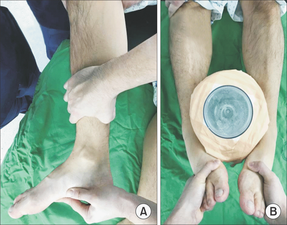
Figure 2.
(A) On anterior drawer radiographs, the degree of anterior translation of talus was defined as the nearest distance between the most posterior portion of tibial plafond and talus. (B) With varus stress radiographs, talar tilting was defined as an angle between articular surfaces of tibial plafond and talus.
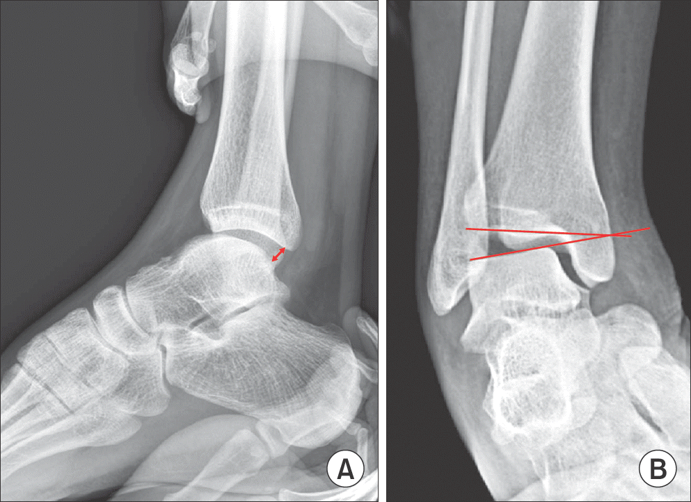
Table 1.
The Result of Initial Sample Test for Consistency of 2 Examiners
Table 2.
The Average Stress Radiographic Values of the Affected Side Related to Anesthesia
Table 3.
Stress Radiographic Values of the Affected Side Subtracted by the Unaffected
 XML Download