Abstract
Tarsal tunnel syndrome is an entrapment neuropathy of the posterior tibial nerve or its branches in the fibro-osseous tunnel beneath the flexor retinaculum. This pathology is associated with multiple etiologies, including trauma, space-occupying lesions, and impaired biomechanics. We report a case of tarsal tunnel syndrome associated with gout tophi in a patient with untreated gout along with a review of the relevant literature on tarsal tunnel syndrome.
Go to : 
REFERENCES
1.Ahmad M., Tsang K., Mackenney PJ., Adedapo AO. Tarsal tunnel syndrome: a literature review. Foot Ankle Surg. 2012. 18:149–52.

2.Lau JT., Daniels TR. Tarsal tunnel syndrome: a review of the lit-erature. Foot Ankle Int. 1999. 20:201–9.

3.Sung KS., Park SJ. Short-term operative outcome of tarsal tunnel syndrome due to benign space-occupying lesions. Foot Ankle Int. 2009. 30:741–5.

4.Lui TH. Acute posterior tarsal tunnel syndrome caused by gouty tophus. Foot Ankle Spec. 2015. 8:320–3.

5.Wakabayashi T., Irie K., Yamanaka H., Iwatani M., Inoue K. Tarsal tunnel syndrome caused by tophaceous gout a case report. J Clin Rheumatol. 1998. 4:151–5.

6.Janssens HJ., Janssen M., van de Lisdonk EH., van Riel PL., van Weel C. Use of oral prednisolone or naproxen for the treatment of gout arthritis: a double-blind, randomised equivalence trial. Lancet. 2008. 371:1854–60.

7.Falidas E., Rallis E., Bournia VK., Mathioulakis S., Pavlakis E., Villias C. Multiarticular chronic tophaceous gout with severe and multiple ulcerations: a case report. J Med Case Rep. 2011. 5:397.

8.Chen CK., Yeh LR., Pan HB., Yang CF., Lu YC., Wang JS, et al. Intra-articular gouty tophi of the knee: CT and MR imaging in 12 patients. Skeletal Radiol. 1999. 28:75–80.

Go to : 
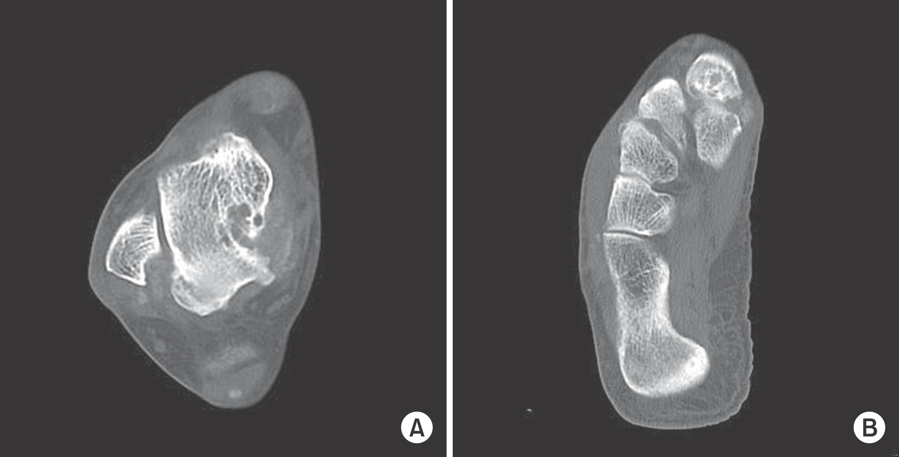
 | Figure 1.Axial computed tomograph shows osteolytic changes with subcortical cysts and multiple calcifications in the talar body (A) and in the first and second tarsometatarsal joints (B). |
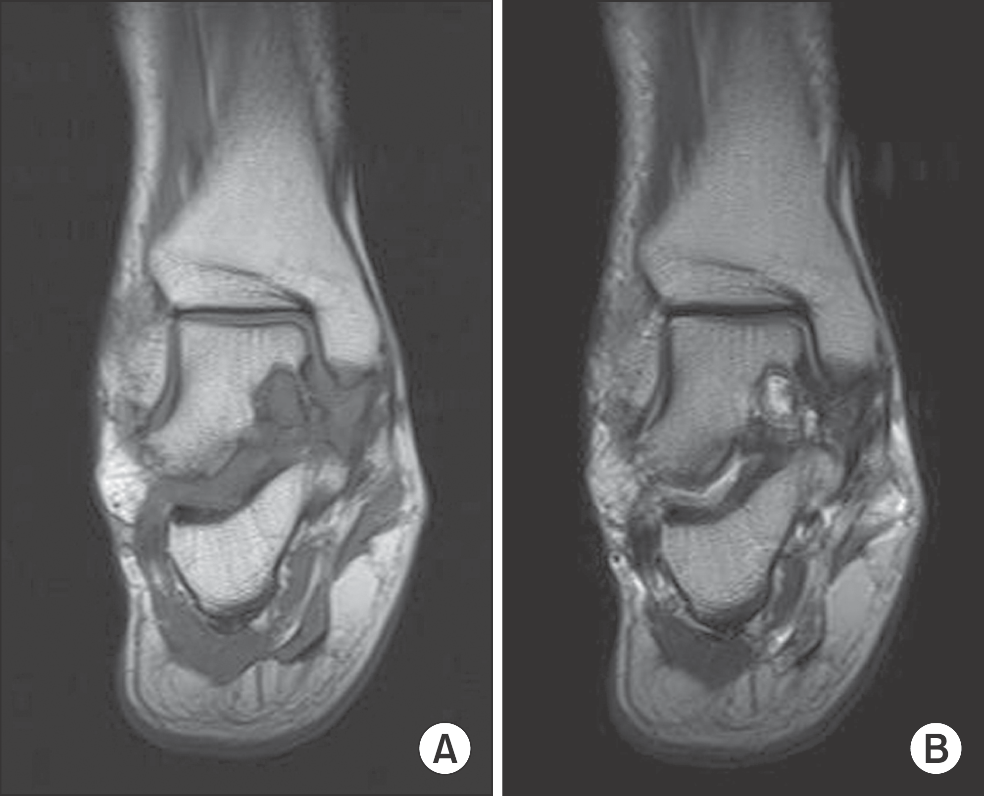
 | Figure 2.Coronal magnetic resonance images show an ill-defined mass in the talar body with low signal intensity on T1-weighted imaging (A) and of heterogeneous signal intensity on T2-weighted imaging (B). |


 PDF
PDF ePub
ePub Citation
 Print
Print


 XML Download