

 PDF
PDF ePub
ePub Citation
 Print
Print


Abstract
Purpose:
This study examined the factors affecting the treatment of diabetes mellitus foot patients who had undergone a Syme amputation.
Materials and Methods:
This study included 17 patients diagnosed with a diabetes mellitus foot and who had undergone a Syme amputation from January 2010 to January 2014. Some of the risk factors (age, body mass index [BMI], disease duration, smoking, ankle brachial index [ABI], HbA1c, serum albumin, total lymphocyte, C-reactive protein [CRP], and serum creatine) that affect the successful Syme amputation were analyzed.
Results:
The healing rate of a Syme amputation was significantly higher when the lymphocyte count was above 1,500 mm3 (p=0.029). The factors affecting the surgical outcome according to multivariate analysis were HbA1c and the BMI (p=0.014, p=0.013). Regarding reamputation, there was a significant difference with HbA1c, lymphocyte, and BMI (p=0.01, p=0.03, and p=0.01). No significant differences were observed with age, disease duration of diabetes mellitus, smoking, ABI, serum albumin, CRP, and serum creatine.
REFERENCES
1.Pinzur MS., Stuck RM., Sage R., Hunt N., Rabinovich Z. Syme ankle disarticulation in patients with diabetes. J Bone Joint Surg Am. 2003. 85:1667–72.

2.Frykberg RG., Abraham S., Tierney E., Hall J. Syme amputation for limb salvage: early experience with 26 cases. J Foot Ankle Surg. 2007. 46:93–100.
3.Dickhaut SC., DeLee JC., Page CP. Nutritional status: importance in predicting wound-healing after amputation. J Bone Joint Surg Am9. 1984. 66:71–5.
4.Wagner FW Jr. Management of the diabetic neurotrophic foot part II. A classification and treatment program for diabetic, neuropathic, and dysvascular foot problems. The American Academy of Orthopaedic Surgeons. editor.Instructional course lectures. St. Louis: CV Mosby;1979. p. 143–65.
5.Syme J. Surgical cases and observations. Amputation at the ankle-joint9 1843. Clin Orthop Relat Res. 1990. 256:3–6.
6.Waters RL., Perry J., Antonelli D., Hislop H. Energy cost of walking of amputees: the influence of level of amputation. J Bone Joint Surg Am. 1976. 58:42–6.
7.Aulivola B., Hile CN., Hamdan AD., Sheahan MG., Veraldi JR., Skillman JJ, et al. Major lower extremity amputation: outcome of a modern series. Arch Surg. 2004. 139:395–9.
8.Yu GV., Schinke TL., Meszaros A. Syme’s amputation: a retrospective review of 10 cases. Clin Podiatr Med Surg. 2005. 22:395–427.
9.Bowker JH. Partial foot and Syme amputations: an overview. Clin Prosthet Orthot. 1987. 12:10–3.
10.Dormandy J., Belcher G., Broos P., Eikelboom B., Laszlo G., Konrad P, et al. Prospective study of 713 below-knee amputations for ischaemia and the effect of a prostacyclin analogue on healing. Hawaii Study Group. Br J Surg. 1994. 81:33–7.
11.Pedersen NW., Pedersen D. Nutrition as a prognostic indicator in amputations. A prospective study of 47 cases. Acta Orthop Scand. 1992. 63:675–8.
12.Christman AL., Selvin E., Margolis DJ., Lazarus GS., Garza LA. Hemoglobin A1c predicts healing rate in diabetic wounds. J Invest Dermatol. 2011. 131:2121–7.
13.Jung HG., Kim YJ., Shim SH., Paik HD. Lower extremity amputations for the diabetic foot complication. J Korean Foot Ankle Soc. 2006. 10:1–6.
14.Reus WF., Robson MC., Zachary L., Heggers JP. Acute effects of tobacco smoking on blood flow in the cutaneous micro-circu-lation. Br J Plast Surg. 1984. 37:213–5.
15.Choi SJ., Lee CB., Kim MS., Ha JH., Park HT. Incidence and risk factors of ipsilateral foot and lower limb reamputation in diabetic foot patients. J Korean Foot Ankle Soc. 2011. 15:7–12.
16.Ohsawa S., Inamori Y., Fukuda K., Hirotuji M. Lower limb amputation for diabetic foot. Arch Orthop Trauma Surg. 2001. 121:186–90.
Figure 1.
(A) A 59-year-old man had type II diabetes, amputated toes and skin necrosis. (B) Syme amputation was performed well and walk freely with prosthesis after healing.
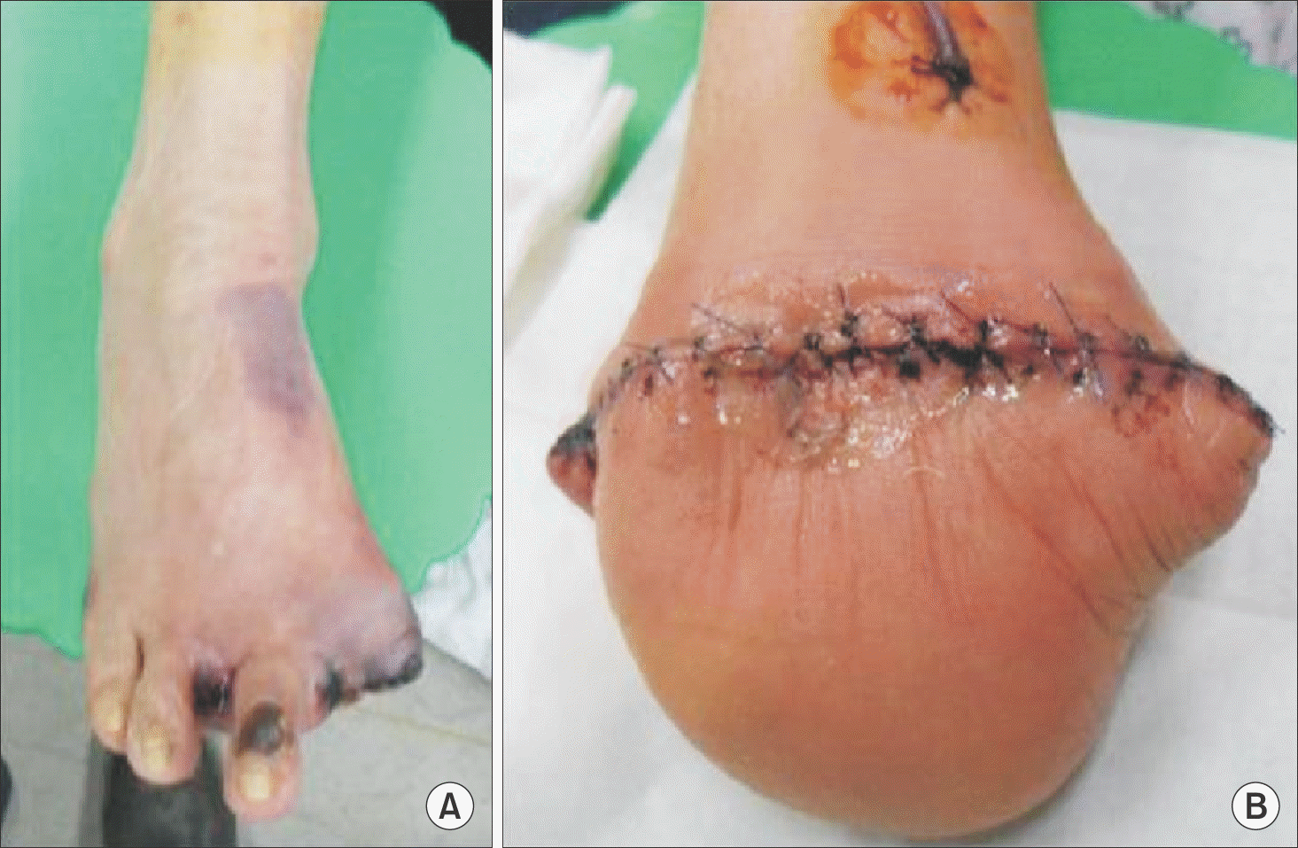
Figure 2.
(A) A 62-year-old man had diabetic foot ulcer. (B) Below knee amputation was performed due to difficulty of infection control and laboratory abnormality.
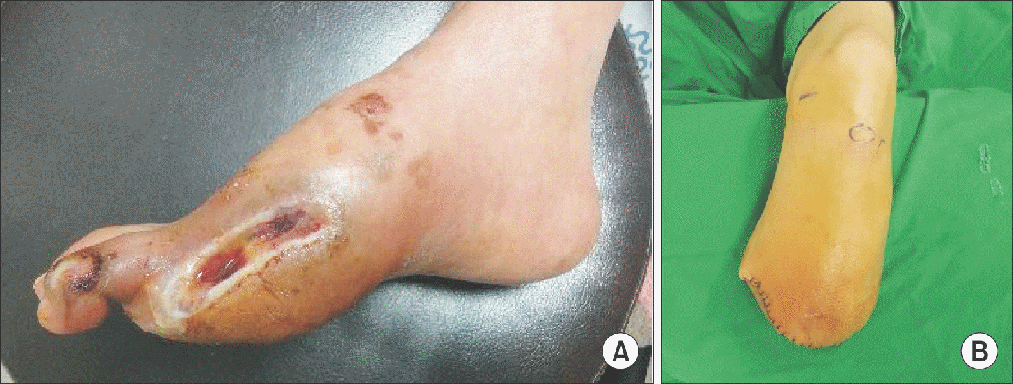
Table 1.
Wound Features in Healed Group and Unhealed Group
| Patient No. | Group | Sex | Age (yr) | Wound site | Size* | Cause |
|---|---|---|---|---|---|---|
| 1 | 1 | Male | 54 | Forefoot | Small | Necrosis |
| 2 | 1 | Male | 50 | Forefoot | Small | Ulcer, infection, necrosis |
| 3 | 1 | Male | 58 | Midfoot | Middle | Necrosis |
| 4 | 1 | Male | 68 | Midfoot | Middle | Infection, necrosis |
| 5 | 1 | Male | 39 | Midfoot | Large | Infection, necrosis |
| 6 | 1 | Male | 57 | Forefoot | Small | Infection, necrosis |
| 7 | 1 | Male | 61 | Forefoot | Small | Necrosis |
| 8 | 1 | Male | 72 | Forefoot | Small | Necrosis |
| 9 | 1 | Male | 70 | Midfoot | Middle | Infection, necrosis |
| 10 | 1 | Female | 78 | Midfoot | Middle | Infection, necrosis |
| 11 | 1 | Male | 85 | Midfoot | Middle | Infection |
| 12 | 1 | Male | 66 | Forefoot | Small | Necrosis |
| 13 | 1 | Female | 46 | Forefoot | Middle | Necrosis |
| 14 | 2 | Female | 49 | Midfoot | Middle | Infection |
| 15 | 2 | Female | 70 | Forefoot | Middle | Ulcer, infection, necrosis |
| 16 | 2 | Male | 62 | Midfoot | Middle | Infection, necrosis |
| 17 | 2 | Female | 85 | Forefoot | Middle | Infection, necrosis |
Table 2.
Rate of Healing by Divided Standard Value
| Factor | No. of patients | Success of treatment (n) | Rate of healing (%) | p-value* | |
|---|---|---|---|---|---|
| Age (yr) | ≥65 | 8 | 6 | 75.0 | 0.665 |
| <65 | 9 | 7 | 77.8 | ||
| HbA1c (%) | ≥8.0 | 12 | 8 | 66.7 | 0.208 |
| <8.0 | 5 | 5 | 100.0 | ||
| Albumin (g/dL) | ≥2.5 | 13 | 11 | 84.6 | 0.219 |
| <2.5 | 4 | 2 | 50.0 | ||
| Smoking | Yes | 9 | 7 | 77.8 | 0.665 |
| No | 8 | 6 | 75.0 | ||
| ABI | ≥0.9 | 4 | 3 | 75.0 | 0.700 |
| <0.9 | 13 | 10 | 76.9 | ||
| BMI (kg/m2) | ≥25.0 | 4 | 2 | 50.0 | 0.219 |
| <25.0 | 13 | 11 | 84.6 | ||
| Creatine (mg/dL) | ≥1.2 | 9 | 7 | 77.7 | 0.665 |
| <1.2 | 8 | 6 | 75.0 | ||
| CRP (mg/L) | ≥3.2 | 13 | 10 | 76.9 | 0.700 |
| <3.2 | 4 | 3 | 75.0 | ||
| Total lymphocyte (mm3) | ≥1,500 | 8 | 8 | 100.0 | 0.029 |
| <1,500 | 9 | 5 | 55.6 | ||
| Wound site | Forefoot | 9 | 7 | 77.7 | 0.893 |
| Midfoot | 8 | 6 | 75.0 | ||
Table 3.
Factors’ Effect to the Operative Outcome
| Factor | p-value* |
|---|---|
| Age | 0.554 |
| DM duration | 0.917 |
| HbA1c | 0.014 |
| Albumin | 0.280 |
| Smoking | 0.663 |
| ABI | 0.841 |
| BMI | 0.013 |
| Creatine | 0.371 |
| CRP | 0.733 |
| Total lymphocyte | 0.052 |
Table 4.
Values of Factors in Healed Group and Unhealed Group
 XML Download