

 PDF
PDF ePub
ePub Citation
 Print
Print


Abstract
Flatfoot deformity, defined as loss of medial longitudinal arch, sometimes involves symptoms such as medial arch pain or Achilles tendon tightening, etc. Whether the etiology of deformity is congenital or acquired, i.e., posterior tibial tendon dysfunction, symptoms are largely resolved with conservative treatment including medication, orthoses, and activity modification. Surgery should be considered in cases of failure of conservative treatment and clinicians can select an appropriate technique among many surgical options including calcaneal osteotomy or flexor digitorum longus tendon transfer. Principles of corrective surgery include the recovery of alignment and the preservation of joint motion.
Go to : 
References
1. Bouchard M, Mosca VS. Flatfoot deformity in children and adolescents: surgical indications and management1 J Am Acad Orthop Surg. 2014; 22:623–32.
2. Johnson KA. Tibialis posterior tendon rupture. Clin Orthop Relat Res. 1983; 177:140–7.

3. Mann RA, Thompson FM. Rupture of the posterior tibial tendon causing flat foot. Surgical treatment. J Bone Joint Surg Am. 1985; 67:556–61.
4. Stein BE, Schon LC. Posterior tibial tendon dysfunction in the adult: current concepts. Instr Course Lect. 2015; 64:441–50.
5. Raikin SM, Winters BS, Daniel JN. The RAM classification: a novel, systematic approach to the adult-acquired flatfoot. Foot Ankle Clin. 2012; 17:169–81.
6. Coughlin MJ, Saltzman CL, Anderson RB. Mann's surgery of the foot and ankle. Philadelphia: Saunders/Elsevier;2014.
7. Marzano R. Nonoperative management of adult flatfoot deformities. Clin Podiatr Med Surg1. 2014; 31:337–47.
8. Sung KS, Yu IS. Acquired adult flatfoot: pathophysiology, diagnosis, and nonoperative treatment. J Korean Foot Ankle Soc. 2014; 18:87–92.
9. Augustin JF, Lin SS, Berberian WS, Johnson JE. Nonoperative treatment of adult acquired flat foot with the Arizona brace. Foot Ankle Clin. 2003; 8:491–502.
10. Wenger DR, Mauldin D, Speck G, Morgan D, Lieber RL. Corrective shoes and inserts as treatment for flexible flatfoot in infants and children. J Bone Joint Surg Am. 1989; 71:800–10.
11. Mann R, Inman VT. Phasic activity of intrinsic muscles of the foot. J Bone Joint Surg Am. 1964; 46:469–81.
12. Alvarez RG, Marini A, Schmitt C, Saltzman CL. Stage I and II posterior tibial tendon dysfunction treated by a structured nonoperative management protocol: an orthosis and exercise program. Foot Ankle Int. 2006; 27:2–8.
13. Kulig K, Burnfield JM, Requejo SM, Sperry M, Terk M. Selective activation of tibialis posterior: evaluation by magnetic resonance imaging. Med Sci Sports Exerc. 2004; 36:862–7.
14. Mosca VS. Calcaneal lengthening for valgus deformity of the hindfoot. Results in children who had severe, symptomatic flatfoot and skewfoot. J Bone Joint Surg Am. 1995; 77:500–12.
15. Mosca VS. Flexible flatfoot and skewfoot. Instr Course Lect. 1996; 45:347–54.
16. Teasdall RD, Johnson KA. Surgical treatment of stage I posterior tibial tendon dysfunction. Foot Ankle Int. 1994; 15:646–8.
17. Crates JM, Richardson EG. Treatment of stage I posterior tibial tendon dysfunction with medial soft tissue procedures. Clin Orthop Relat Res. 1999; 365:46–9.
18. Kwon JY, Myerson MS. Management of the flexible flat foot in the child: a focus on the use of osteotomies for correction. Foot Ankle Clin. 2010; 15:309–221.
19. Nyska M, Parks BG, Chu IT, Myerson MS. The contribution of the medial calcaneal osteotomy to the correction of flatfoot deformities. Foot Ankle Int. 2001; 22:278–82.
20. Sung IH, Lee S, Otis JC, Deland JT. Posterior tibial tendon force requirement in early heel rise after calcaneal osteotomies. Foot Ankle Int. 2002; 23:842–9.
21. Evans D. Calcaneo-valgus deformity. J Bone Joint Surg Br. 1975; 57:270–8.
22. Sangeorzan BJ, Mosca V, Hansen ST Jr. Effect of calcaneal lengthening on relationships among the hindfoot, midfoot, and forefoot. Foot Ankle1. 1993; 14:136–41.
23. Ellis SJ, Williams BR, Garg R, Campbell G, Pavlov H, Deland JT. Incidence of plantar lateral foot pain before and after the use of trial metal wedges in lateral column lengthening. Foot Ankle Int. 2011; 32:665–73.
24. Gutiérrez PR, Lara MH. Giannini prosthesis for flatfoot. Foot Ankle Int. 2005; 26:918–26.
25. Moon JS, Bae WH, Seo JG, Lee WC. Clinical results of the subtalar arthroereisis for the flat foot. J Korean Foot Ankle Soc. 2008; 12:117–21.
26. Needleman RL. A surgical approach for flexible flatfeet in adults including a subtalar arthroereisis with the MBA sinus tarsi implant. Foot Ankle Int. 2006; 27:9–18.
Go to :
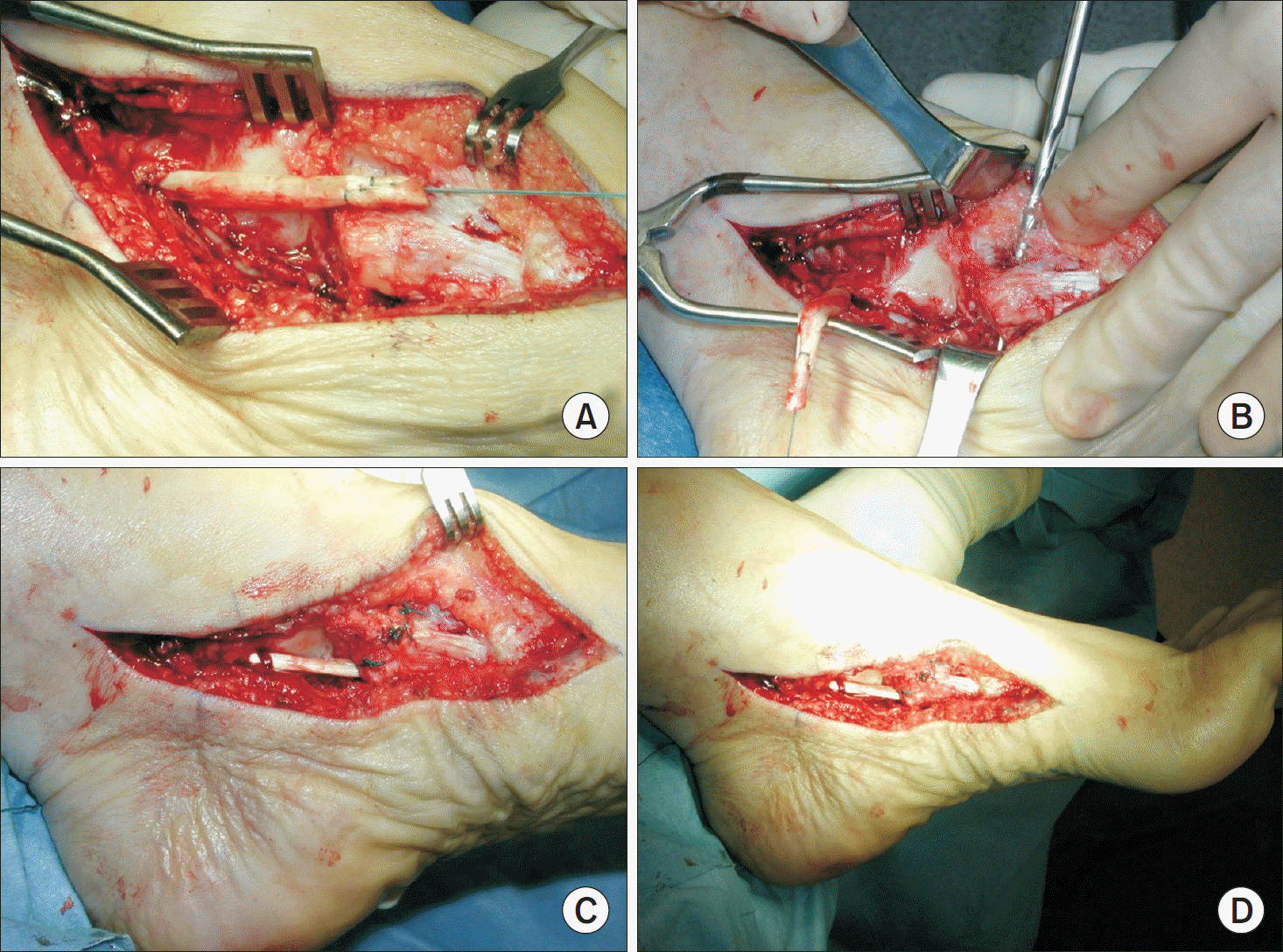 | Figure 1.Flexor digitorum longus (FDL) transfer procedure. (A) Photograph shows isolated FDL tendon. Drilling at the navicular tuberosity (B), then tenodesis is performed Fig. 1C and D. |
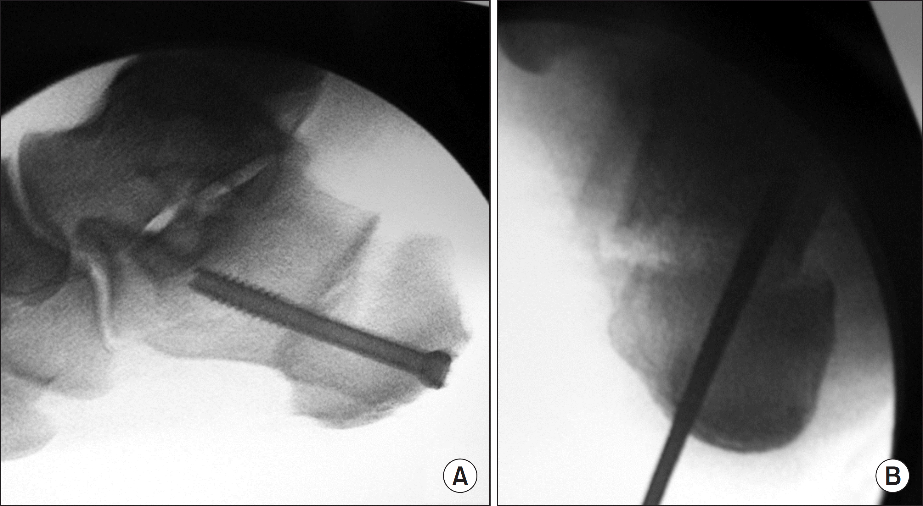 | Figure 2.Fluoroscopy shows medial sliding calcaneal osteotomy at sagittal view (A) and axial view (B). |
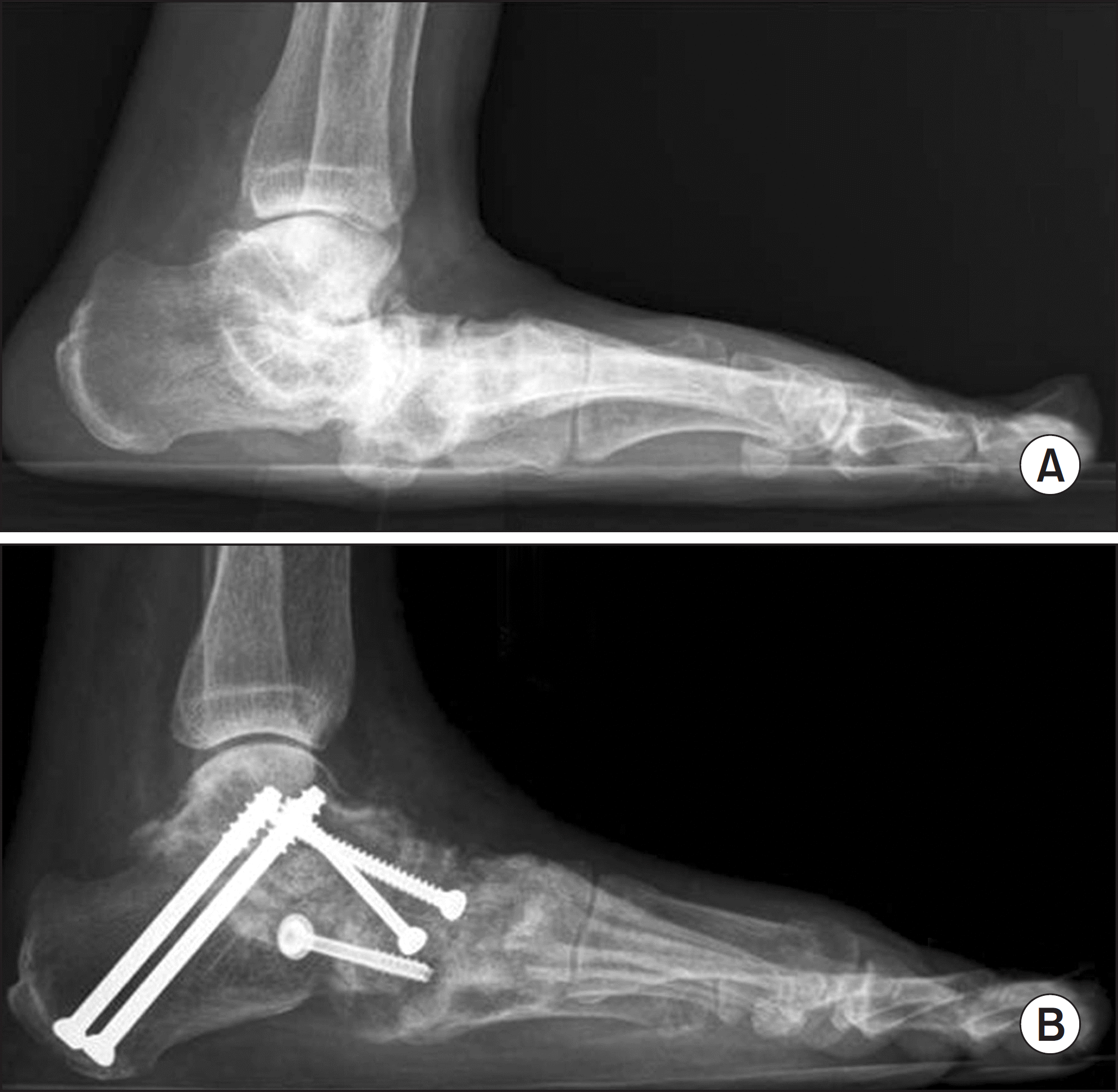
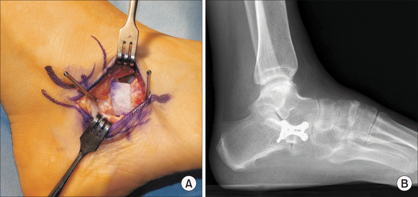 | Figure 3.(A) Interpositional allo-bone graft was performed at calcaneal anterior process. (B) Postoperative lateral standing radiograph shows elevated calcaneal pitch after lateral column lengthening. |
 XML Download