

 PDF
PDF ePub
ePub Citation
 Print
Print


Abstract
The cavus foot is a deformity characterized by an elevated medial longitudinal arch and a hindfoot varus with plantarflexed 1st ray. The etiology of cavus foot is usually related to neuromuscular disease or idiopathic cause. Thorough clinical and radiographic evaluation is required for differentiating etiology of the cavus. Most cases of cavus foot are stable and slowly progressive deformities which can initially be managed with conservative treatment including orthoses and physical therapies. Determining whether the deformity is flexible or rigid, the apex of the deformity and any muscle imbalances in foot and ankle is important for achievement of an adequately balanced plantigrade foot. Treatment should include systematic preoperative planning for selection of appropriate procedures for maintaining a functional and flexible foot with combinations of soft-tissue release, osteotomy, tendon transfer, and arthrodesis.
REFERENCES
1.Faldini C., Traina F., Nanni M., Mazzotti A., Calamelli C., Fabbri D, et al. Surgical treatment of cavus foot in Charcot-Marie-Tooth disease: a review of twenty-four cases: AAOS exhibit selection. J Bone Joint Surg Am. 2015. 97:e30.
2.Fabian G., Krause GPG. Pes cavus. Coughlin MJ, Saltzman CL, Anderson RB, editors. editors.Mann’s surgery of the foot and ankle. 9th ed.Philadelphia: Saunders/Elsevier;20149. p91361-82.
3.Nogueira MP., Farcetta F., Zuccon A. Cavus foot. Foot Ankle Clin. 2015. 20:645–56.

4.Ortiz C., Wagner E., Keller A. Cavovarus foot reconstruction. Foot Ankle Clin. 2009. 14:471–87.
5.Brewerton DA., Sandifer PH., Sweetnam DR. “Idiopathic” pes cavus: an investigation into its aetiology. Br Med J. 1963. 2:659–61.
6.Holmes JR., Hansen ST Jr. Foot and ankle manifestations of Charcot-Marie-Tooth disease. Foot Ankle. 1993. 14:476–86.
7.Nagai MK., Chan G., Guille JT., Kumar SJ., Scavina M., Mackenzie WG. Prevalence of Charcot-Marie-Tooth disease in patients who have bilateral cavovarus feet. J Pediatr Orthop. 2006. 26:438–439.
8.Marks RM. Midfoot and forefoot issues cavovarus foot: assessment and treatment issues9 Foot Ankle Clin. 2008. 13:229–41.
9.Deben SE., Pomeroy GC. Subtle cavus foot: diagnosis and man-agement. J Am Acad Orthop Surg. 2014. 22:512–20.
10.Manoli A 2nd., Graham B. The subtle cavus foot, “the underpro-nator”. Foot Ankle Int. 2005. 26:256–63.
11.Manoli A 2nd., Smith DG., Hansen ST Jr. Scarred muscle excision for the treatment of established ischemic contracture of the lower extremity. Clin Orthop Relat Res. 1993. 292:309–14.
12.Abbasian A., Pomeroy G. The idiopathic cavus foot-not so subtle after all. Foot Ankle Clin. 2013. 18:629–42.
13.Mann RA., Missirian J. Pathophysiology of Charcot-Marie-Tooth disease. Clin Orthop Relat Res. 1988. 234:221–8.
14.Mosca VS. The cavus foot. J Pediatr Orthop. 2001. 21:423–4.
15.Aminian A., Sangeorzan BJ. The anatomy of cavus foot deformity. Foot Ankle Clin. 2008. 13:191–8.
16.Martyn CN., Hughes RA. Epidemiology of peripheral neuropathy. J Neurol Neurosurg Psychiatry. 1997. 62:310–8.
17.Guyton GP., Mann RA. The pathogenesis and surgical management of foot deformity in Charcot-Marie-Tooth disease. Foot Ankle Clin. 2000. 5:317–26.
18.VanderHave KL., Hensinger RN., King BW. Flexible cavovarus foot in children and adolescents. Foot Ankle Clin. 2013. 18:715–26.
19.Lee MC., Sucato DJ. Pediatric issues with cavovarus foot defor-mities. Foot Ankle Clin. 2008. 13:199–219.
20.Tynan MC., Klenerman L., Helliwell TR., Edwards RH., Hayward M. Investigation of muscle imbalance in the leg in symptomatic forefoot pes cavus: a multidisciplinary study. Foot Ankle. 1992. 13:489–501.
21.Wicart P. Cavus foot, from neonates to adolescents. Orthop Traumatol Surg Res. 2012. 98:813–28.
22.Barenfeld PA., Weseley MS., Shea JM. The congenital cavus foot. Clin Orthop Relat Res. 1971. 79:119–26.
23.Perera A., Guha A. Clinical and radiographic evaluation of the cavus foot: surgical implications. Foot Ankle Clin. 2013. 18:619–28.
24.Saltzman CL., el-Khoury GY. The hindfoot alignment view. Foot Ankle Int. 1995. 16:572–6.
25.Alexander IJ., Johnson KA. Assessment and management of pes cavus in Charcot-Marie-Tooth disease. Clin Orthop Relat Res. 1989. 246:273–81.
26.Dwyer FC. The present status of the problem of pes cavus. Clin Orthop Relat Res. 1975. 106:254–75.
27.Georgiadis AG., Spiegel DA., Baldwin KD. The cavovarus foot in hereditary motor and sensory neuropathies. JBJS Reviews. 2015. 3:e5.
28.Mubarak SJ., Van Valin SE. Osteotomies of the foot for cavus deformities in children. J Pediatr Orthop. 2009. 29:294–9.
29.Aktas S., Sussman MD. The radiological analysis of pes cavus deformity in Charcot Marie Tooth disease. J Pediatr Orthop B. 2000. 9:137–40.
30.Bradley GW., Coleman SS. Treatment of the calcaneocavus foot deformity. J Bone Joint Surg Am. 1981. 63:1159–66.
33.Jahss MH. Tarsometatarsal truncated-wedge arthrodesis for pes cavus and equinovarus deformity of the fore part of the foot. J Bone Joint Surg Am. 1980. 62:713–22.
34.Cole WH. The treatment of claw-foot. J Bone Joint Surg Am. 1940. 22:895–908.
35.Japas LM. Surgical treatment of pes cavus by tarsal V-osteotomy. Preliminary report. J Bone Joint Surg Am. 1968. 50:927–44.
36.Dwyer FC. Osteotomy of the calcaneum for pes cavus. J Bone Joint Surg Br. 1959. 41:80–6.
37.Knupp M., Horisberger M., Hintermann B. A new z-shaped calcaneal osteotomy for 3-plane correction of severe varus deformity of the hindfoot. Tech Foot Ankle Surg. 2008. 7:90–5.
38.Koutsogiannis E. Treatment of mobile flat foot by displacement osteotomy of the calcaneus. J Bone Joint Surg Br. 1971. 53:96–100.
39.Krause F., Windolf M., Schwieger K., Weber M. Ankle joint pressure in pes cavovarus. J Bone Joint Surg Br. 2007. 89:1660–5.
40.Bariteau JT., Blankenhorn BD., Tofte JN., DiGiovanni CW. What is the role and limit of calcaneal osteotomy in the cavovarus foot? Foot Ankle Clin. 2013. 18:697–714.
41.Kaplan JR., Myerson MS. The failed cavovarus foot: what went wrong and why? Instr Course Lect. 2016. 65:331–44.
42.Henderson CP., Parks BG., Guyton GP. Lateral and medial plantar pressures after split versus whole anterior tibialis tendon trans-fer. Foot Ankle Int. 2008. 29:1038–41.
43.Ward CM., Dolan LA., Bennett DL., Morcuende JA., Cooper RR. Long-term results of reconstruction for treatment of a flexible cavovarus foot in Charcot-Marie-Tooth disease. J Bone Joint Surg Am. 2008. 90:2631–42.
Figure 1.
This figure shows Coleman block test of right foot for assessing flexibility of hindfoot varus. By weight-bearing on a block supporting only the lateral side of the foot except 1st ray, if the hindfoot remains flexible, the hindfoot varus can be corrected by addressing the forefoot deformity alone.

Figure 2.
On standing lateral view of cavus foot, (a) talo-1st metatarsal angle (Meary’s angle), (b) calcaneal pitch angle, and (c) calcaneo-1st metatarsal angle (Hibb’s angle) can be measured.
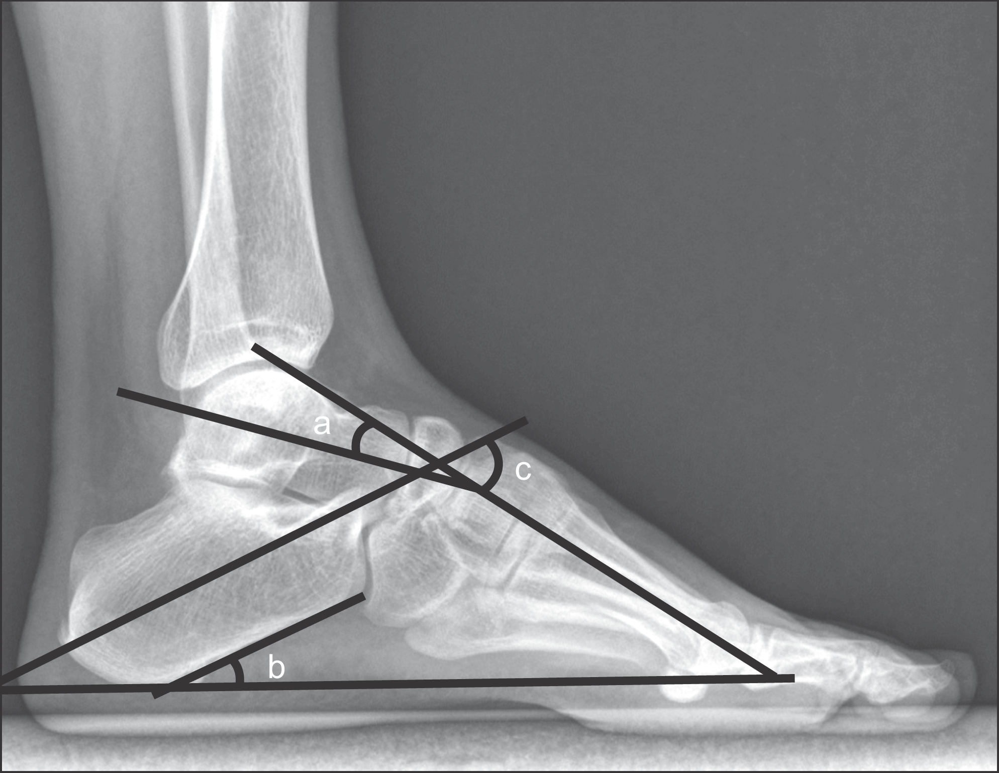
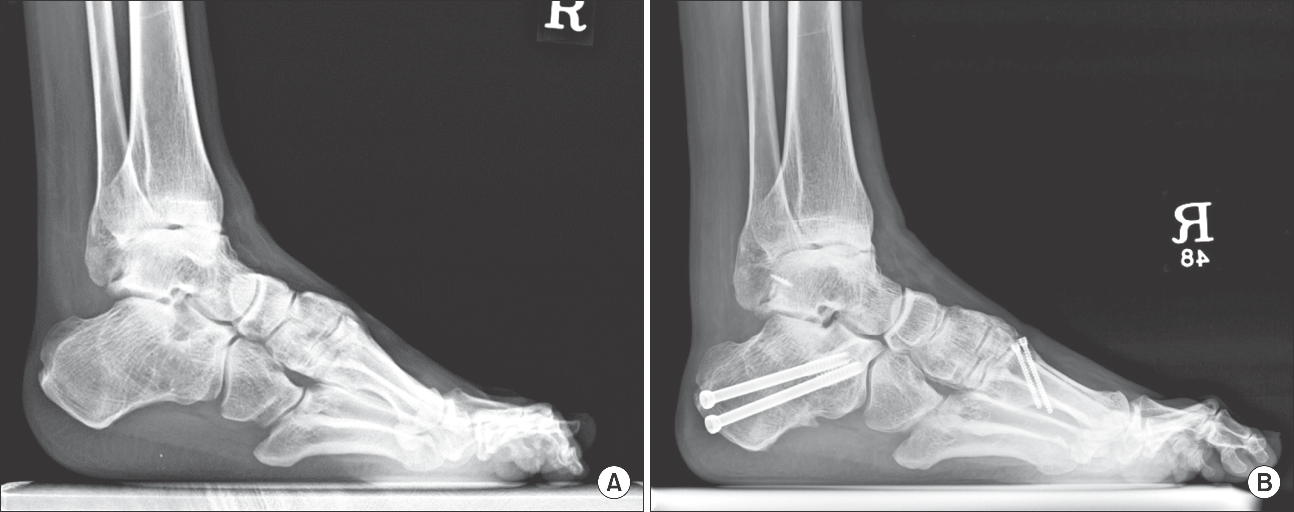
Figure 3.
(A) The standing lateral radiograph of right foot showed a severe cavovarus deformity. Reconstruction of cavovarus foot was done with a calcaneal lateral closing wedge osteotomy, 1st metatarsal dorsiflexion osteotomy, plantar fascia release, Achilles tendon lengthening, a modified Broström operation and tibialis anterior tendon lateralization. (B) The standing lateral radiograph for 1-year follow-up after surgery showed a balanced and plantigrade foot.
 XML Download