Abstract
Development of a pseudoaneurysm around the ankle is an uncommon complication after surgery. We experienced a case of a pseudoaneurysm, which developed from the anterior tibial artery. A 44-year-old woman had sustained painful swelling of her right ankle after the removal of implants for a distal fibular fracture. The pseudoaneurysm was confirmed by ultrasonography and angiography. The patient was treated with an intervention using a coil and recovered without further complaints. This case report aims to increase the awareness of this complication with review of literature.
References
1. Yu JL, Ho E, Wines AP. Pseudoaneurysms around the foot and ankle: case report and literature review. Foot Ankle Surg. 2013; 19:194–8.

2. Chun TH, Park YS, Kim YT, Sung KS. Pseudoaneurysm of anterior tibial artery after ankle arthroscopy. J Korean Foot Ankle Soc. 2012; 16:265–9.
3. Baek JR, Park HK, Yang SH. Pseudoaneurysm of the anterior tibial artery: a case report. J Korean Foot Ankle Soc. 2007; 11:104–6.
4. Han KJ, Won YY, Kim TY, Khang SY. Pseudoaneurysm of the anterior tibial artery after closed intramedullary nailing of a tibial shaft fracture: a case report. J Korean Orthop Assoc. 2002; 37:574–6.

5. Kim TS, Whang KS, Seo WY. Traumatic pseudoaneurysm of posterior tibial artery in a child: a case report. J Korean Fract Soc. 2007; 20:83–5.

6. Mariani PP, Mancini L, Giorgini TL. Pseudoaneurysm as a complication of ankle arthroscopy. Arthroscopy. 2001; 17:400–2.

7. Matson MB, Morgan RA, Belli AM. Percutaneous treatment of pseudoaneurysms using fibrin adhesive. Br J Radiol. 2001; 74:690–4.

8. McIvor J, Treweeke PS. Direct percutaneous embolisation of a false aneurysm with steel coils. Clin Radiol. 1988; 39:205–7.
Figure 1.
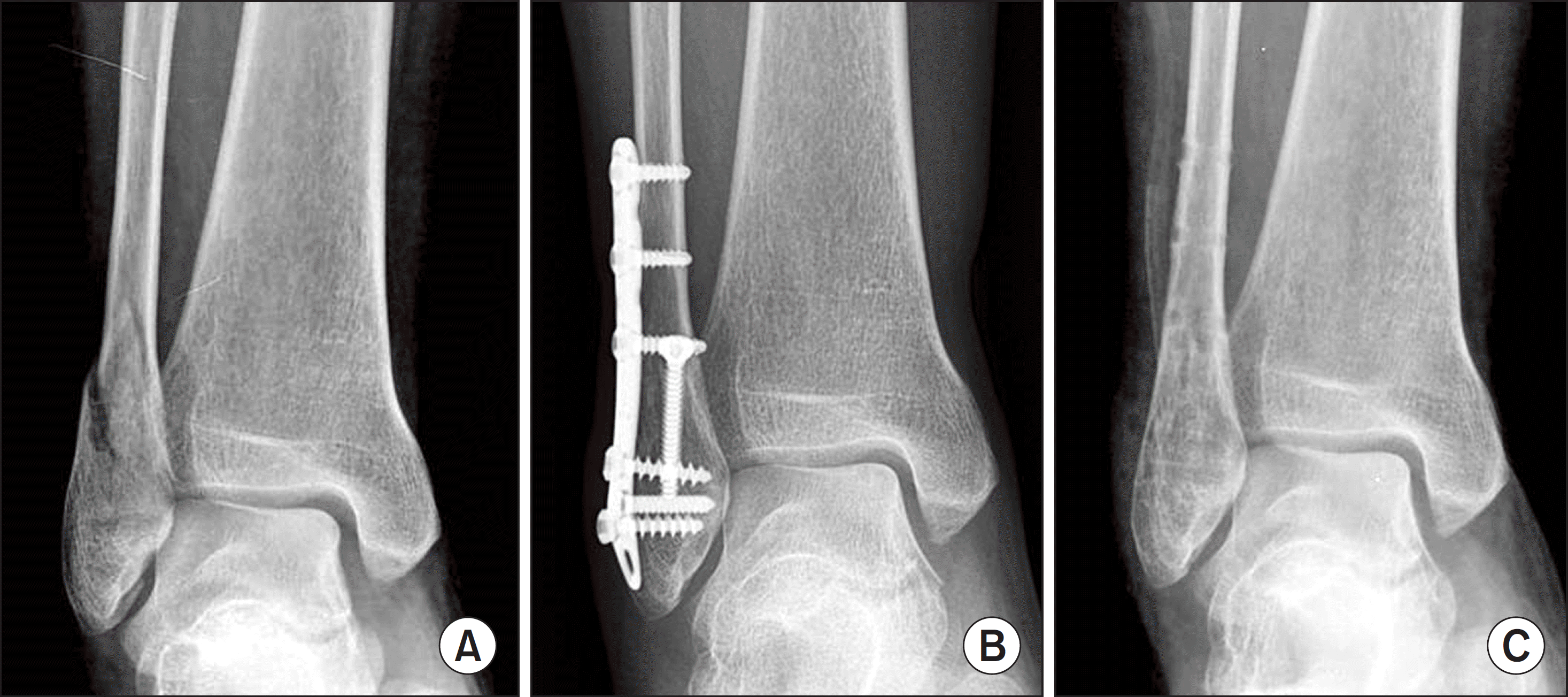
(A) Initial anteroposterior radiograph demonstrates the distal fibular fracture. (B) Follow-up radiograph at postoperative 12 months after initial operation demonstrates the bone union. (C) Postoperative radiograph demonstrates the removal status of metal implants.

Figure 2.
(A) Ultrasonography shows hypoechoic oval shaped lesion beside the fibula. (B) Color flow Doppler scan shows blood circulation into the pseudoaneurysm.

Figure 3.
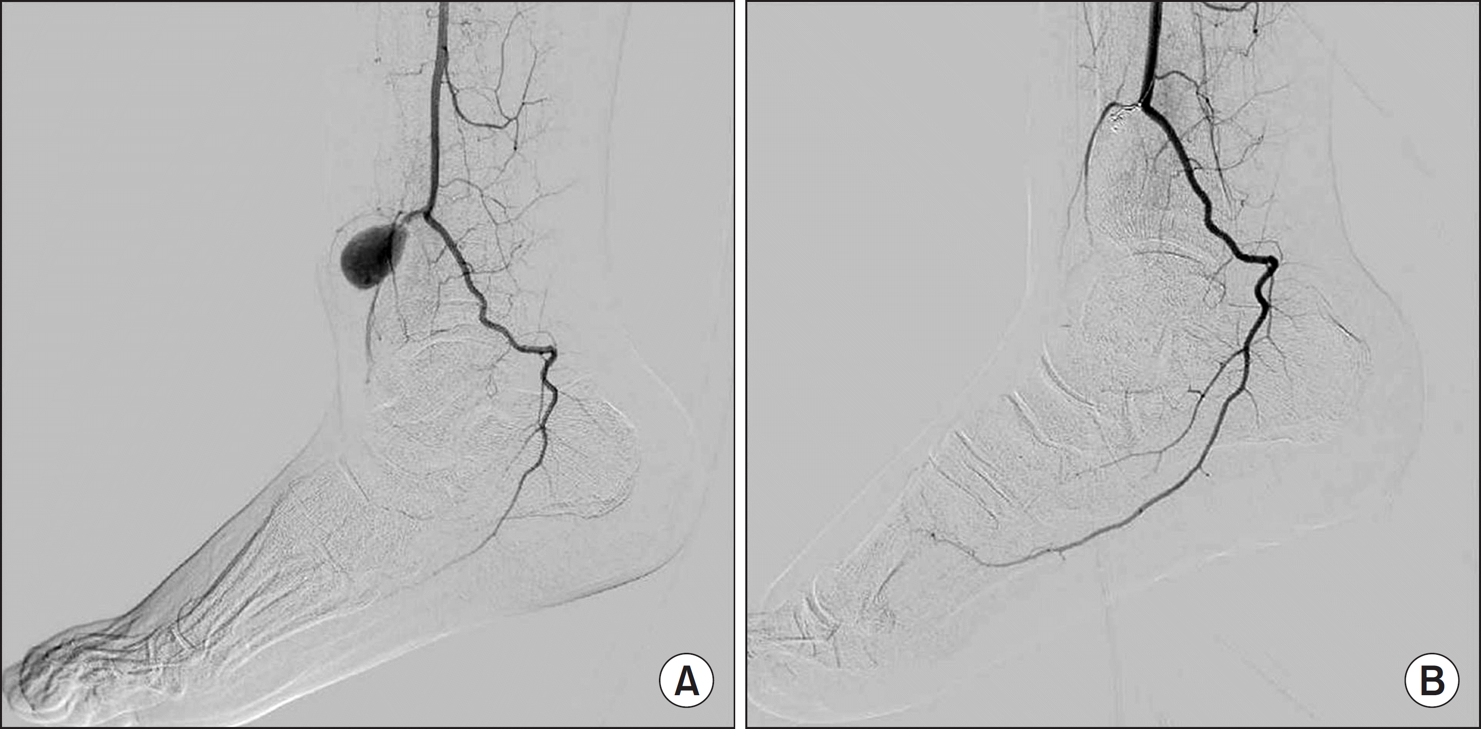
(A) Femoral arteriogram shows pseudoaneurysm of anterior tibial artery. (B) Pseudoaneurysm completely excluded from circulation by embolization with microcoils.

Table 1.
Review of Reported Cases in the Korean Literature


 PDF
PDF ePub
ePub Citation
 Print
Print


 XML Download