Abstract
The tibialis anterior tendon functions as a major dorsiflexor of the ankle. A rupture in this tendon can cause serious problems in the ambulatory function. A closed traumatic rupture without open wound or an atraumatic rupture can delay diagnosis and treatment. There are not enough guidelines for an effective surgical treatment on this chronic condition. Herein, we report two cases of chronic tibialis anterior disruption successfully treated by semitendinosus autograft.
Go to : 
REFERENCES
1.Cornwall MW., McPoil TG. The influence of tibialis anterior muscle activity on rearfoot motion during walking. Foot Ankle Int. 1994. 15:75–9.

2.Markarian GG., Kelikian AS., Brage M., Trainor T., Dias L. Anterior tibialis tendon ruptures: an outcome analysis of operative versus nonoperative treatment. Foot Ankle Int. 1998. 19:792–802.

3.van Acker G., Pingen F., Luitse J., Goslings C. Rupture of the tibialis anterior tendon. Acta Orthop Belg. 2006. 72:105–7.
4.Sammarco VJ., Sammarco GJ., Henning C., Chaim S. Surgical repair of acute and chronic tibialis anterior tendon ruptures. J Bone Joint Surg Am. 2009. 91:325–32.
5.Kausch T., Rütt J. Subcutaneous rupture of the tibialis anterior tendon: review of the literature and a case report. Arch Orthop Trauma Surg. 1998. 117:290–3.

6.Wong MW. Traumatic tibialis anterior tendon rupture-delayed repair with free sliding tibialis anterior tendon graft. Injury. 2004. 35:940–4.

7.Grundy JR., O’Sullivan RM., Beischer AD. Operative management of distal tibialis anterior tendinopathy. Foot Ankle Int. 2010. 31:212–9.

8.Michels F., Van Der Bauwhede J., Oosterlinck D., Thomas S., Guillo S. Minimally invasive repair of the tibialis anterior tendon using a semitendinosus autograft. Foot Ankle Int. 2014. 35:264–71.

Go to : 
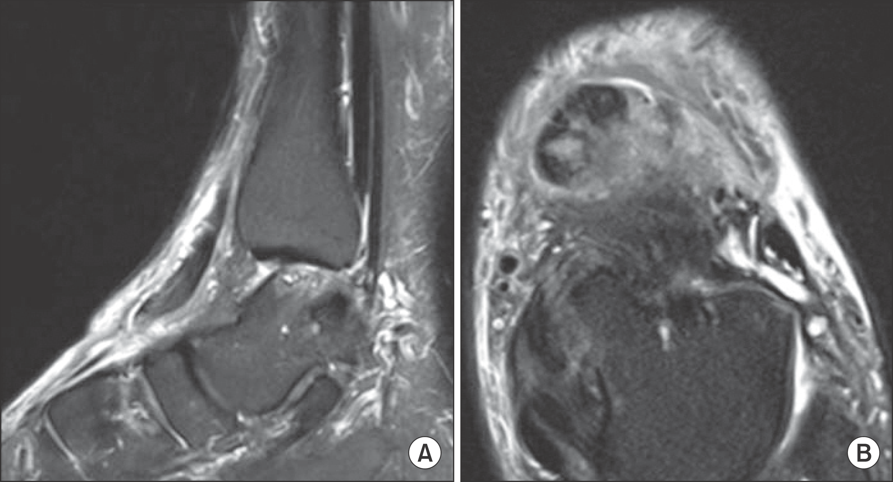 | Figure 1.(A) Sagittal view of magnetic resonance imaging (MRI) demonstrates complete ruptured tibialis anterior tendon retracted at the talonavicular joint level. (B) Axial view of MRI demonstrates proximal tendon stump formed a fibrotic lump. |
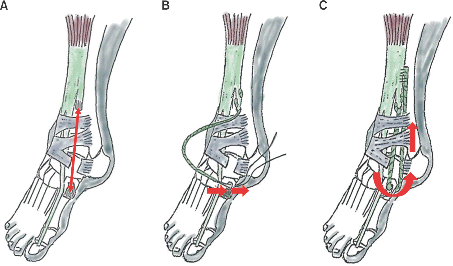 | Figure 3.(A) The final defect length was measured. (B) Semintendinosus autograft was sutured to the proximal stump by Pulvertaft weave and the distal end was passed through the bone tunnel of the medial cuneiform. (C) Distal end of the autograft was pulled back proximally and was sutured to the normal tendinous portion of the proximal stump. |
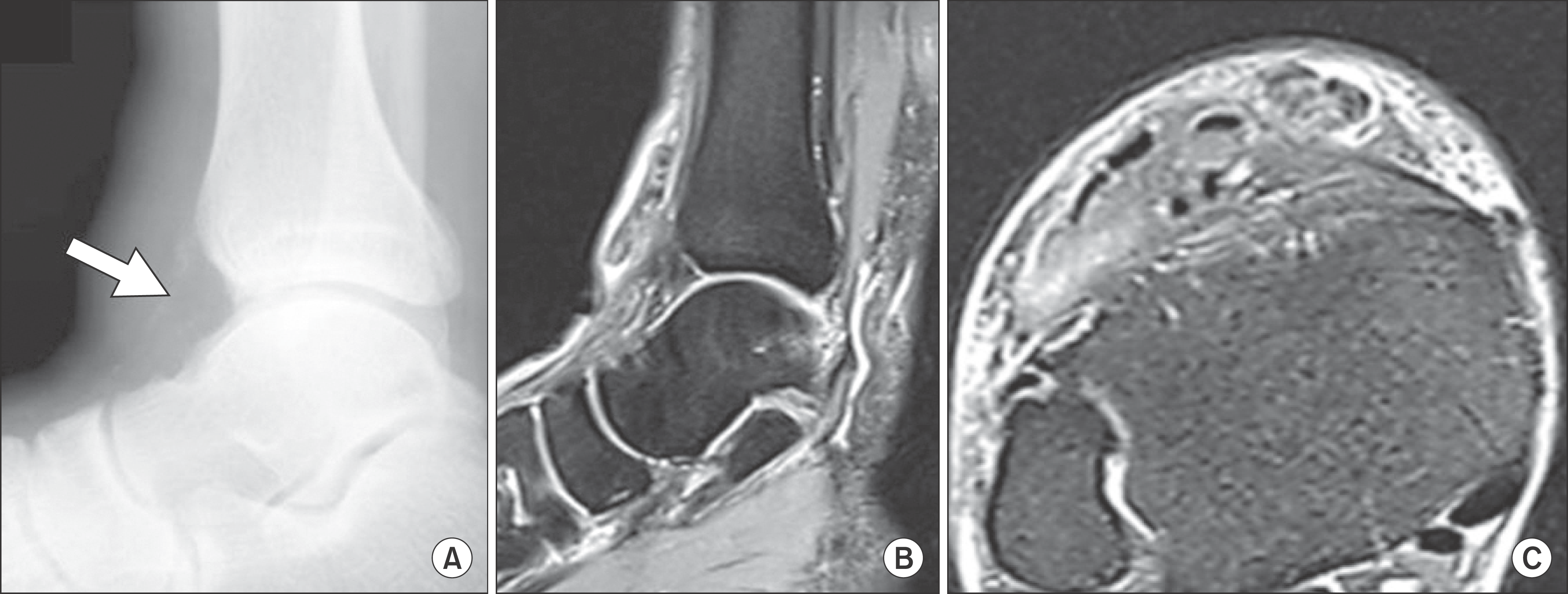 | Figure 4.(A) Lateral radiograph of the ankle shows calcification deposit on the anterior aspect of the tibiotalar joint (arrow). (B) A sagittal view of magnetic resonance image (MRI) demonstrates completely ruptured tibialis anterior tendon with retraction. (C) An axial view of MRI demonstrates proximal stump of the ruptured tibialis anterior tendon forming a fibrotic lump. |


 PDF
PDF ePub
ePub Citation
 Print
Print


 XML Download