Abstract
Osteochondroma is one of the most common bone tumors. It can occur anywhere, although it is most frequent mainly around the metaphysis of long bones. Prediction sites are distal femur, proximal humerus, proximal tibia, and so on. However, osteochondroma in sesamoid is very rare. Herein, we report a case of a 56-year-old woman with symptomatic extra-articular osteochondroma in hallucal sesamoid with a brief literature review.
REFERENCES
1.Giudici MA., Moser RP Jr., Kransdorf MJ. Cartilaginous bone tu-mors. Radiol Clin North Am. 1993. 31:237–59.
2.Torreggiani WC., Munk PL., Al-Ismail K., O’Connell JX., Nicolaou S., Lee MJ, et al. MR imaging features of bizarre parosteal osteochondromatous proliferation of bone (Nora’s lesion). Eur J Radiol. 2001. 40:224–31.

3.Greger G., Catanzariti AR. Osteochondroma: review of the lit-erature and case report. J Foot Surg. 1992. 31:298–300.
4.Karasick D., Schweitzer ME., Eschelman DJ. Symptomatic osteochondromas: imaging features. AJR Am J Roentgenol. 1997. 168:1507–12.

5.Mowad SC Sr., Zichichi S., Mullin R. Osteochondroma of the tibial sesamoid. J Am Podiatr Med Assoc. 1995. 85:765–6.

6.Okada A., Hatori M., Hashimoto Y., Lee E. Painful extraskeletal osteochondroma under the tarsal sesamoid: a case report and review of literature. Eur J Orthop Surg Traumatol. 2012. 22(Suppl 1):215-220.

7.Nora FE., Dahlin DC., Beabout JW. Bizarre parosteal osteochondromatous proliferations of the hands and feet Am J Surg Pathol. 1983. 7:245–50.
Figure 1.
(A, B) Clinical photographs of preoperative foot status show that a corn on the first metatarsal head area was occurred due to bony protrusion.
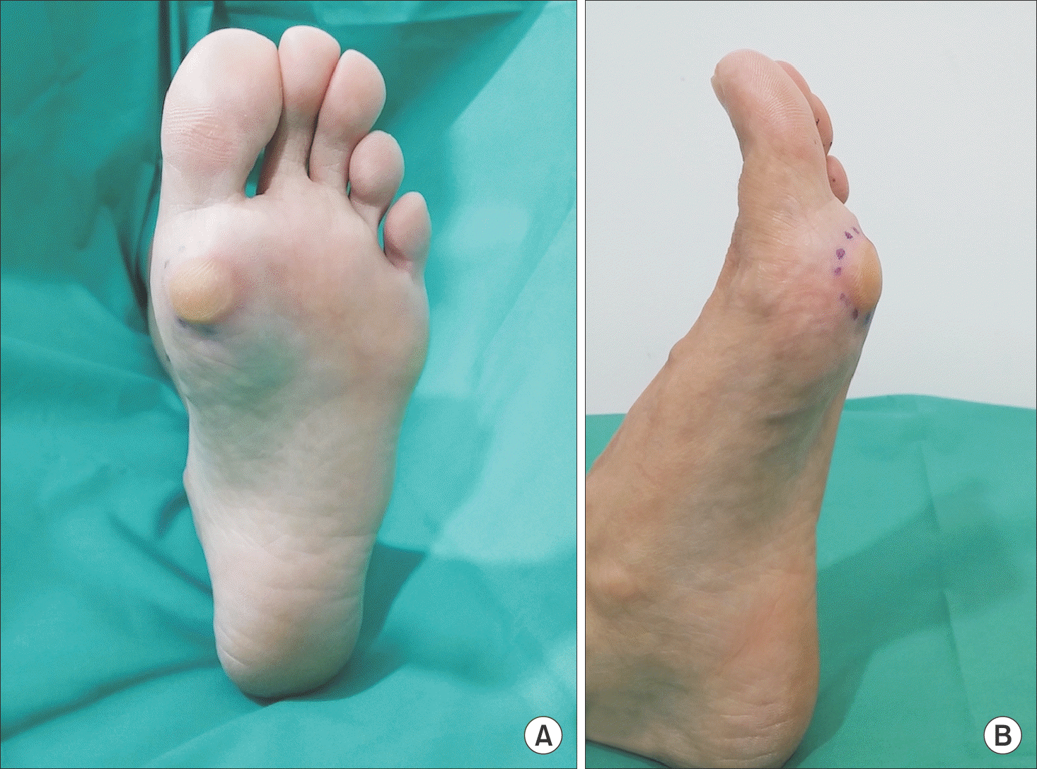
Figure 2.
(A, B) Preoperative radiographs of both feet show a bony mass of tibial sesamoid on big toe of the left foot in the anteroposterior and tangential views (arrows).
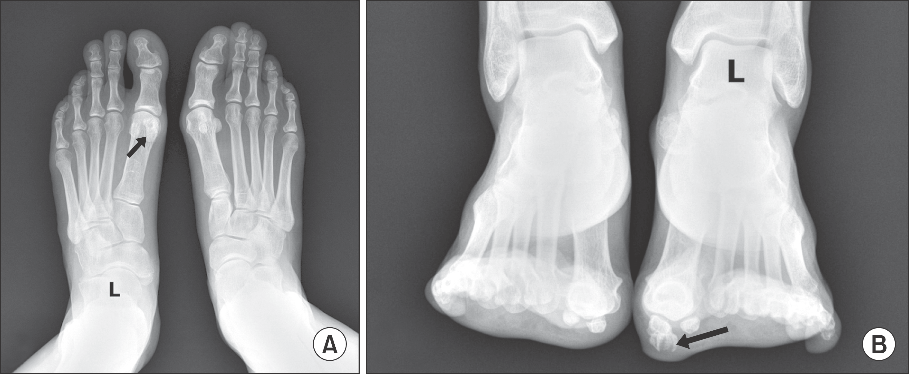
Figure 3.
Axial image (A) and sagittal image (B) of the left foot computed tomographs demonstrate 10×11×8 mm3 sized bony mass with irregular margin from the tibial sesamoid (arrows).

Figure 4.
On the magnetic resonance images, axial view (A) and sagittal view (C) of T1-weighted image show low signal intensity of bony mass (arrows). Axial view (B) and sagittal view (D) of T2-weighted image also show low signal intensity of bony mass (arrows).


 PDF
PDF ePub
ePub Citation
 Print
Print


 XML Download