

 PDF
PDF ePub
ePub Citation
 Print
Print


Abstract
Purpose:
Tarsal coalition results from defects during the developmental stage and produes ankle pain and limitations in the range of motions. Its incidence has been reported to be 1%, but there has not been any reports with respect to Koreans. Therefore, we evaluated the prevalence of tarsal coalition in Koreans.
Materials and Methods:
Between 2005 and 2014, we analyzed a total of 733 cases of foot and ankle magnetic resonance imaging (MRI) in our hospital. There were 391 men and 342 women. All MRI readings were read by a radiologist in our hospital. We classified the coalitions in accordance with the histological and anatomical characteristics, and calculated the prevalence in each group. Moreover, we tried to determine the prevalence of tarsal coalitions in accordance with sex, age, and proportion of the symptomatic tarsal coalitions.
Go to : 
REFERENCES
1.Harris BJ. Anomalous structure in the developing human foot. Anat Rec. 1955. 121:399.
2.Jack EA. Bone anomalies of the tarsus in relation to peroneal spastic flat foot. J Bone Joint Surg Br. 1954. 36:532–42.

3.Jayakumar S., Cowell HR. Rigid flatfoot. Clin Orthop Relat Res. 1977. 122:77–84.
4.Mosier KM., Asher M. Tarsal coalitions and peroneal spastic flat foot. A review. J Bone Joint Surg Am. 1984. 66:976–84.
5.Zaw H., Calder JD. Tarsal coalitions. Foot Ankle Clin. 2010. 15:349–64.
6.Nalaboff KM., Schweitzer ME. MRI of tarsal coalition: frequency, distribution, and innovative signs. Bull NYU Hosp Jt Dis. 2008. 66:14–21.
7.Newman JS., Newberg AH. Congenital tarsal coalition: multimodality evaluation with emphasis on CT and MR imaging. Radio-graphics. 2000. 20:321–32.
8.Wechsler RJ., Schweitzer ME., Deely DM., Horn BD., Pizzutillo PD. Tarsal coalition: depiction and characterization with CT and MR imaging. Radiology. 1994. 193:447–52.
9.Pachuda NM., Lasday SD., Jay RM. Tarsal coalition: etiology, diagnosis, and treatment. J Foot Surg. 1990. 29:474–88.
10.Harris RI., Beath T. Etiology of peroneal spastic flat foot. J Bone Joint Surg Br. 1948. 30:624–34.
11.Varner KE., Michelson JD. Tarsal coalition in adults1 Foot Ankle Int1. 2000. 21:669–72.
12.Conway JJ., Cowell HR. Tarsal coalition: clinical significance and roentgenographic demonstration. Radiology. 1969. 92:799–811.
13.Munk PL., Vellet AD., Levin MF., Helms CA. Current status of magnetic resonance imaging of the ankle and the hindfoot. Can Assoc Radiol J. 1992. 43:19–30.
14.Masciocchi C., D’Archivio C., Barile A., Fascetti E., Zobel BB., Gallucci M, et al. Talocalcaneal coalition: computed tomography and magnetic resonance imaging diagnosis. Eur J Radiol. 1992. 15:22–5.
15.Leonard MA. The inheritance of tarsal coalition and its relationship to spastic flat foot. J Bone Joint Surg Br. 1974. 56:520–6.
16.Stormont DM., Peterson HA. The relative incidence of tarsal co-alition. Clin Orthop Relat Res. 1983. 181:28–36.
17.Cowell HR., Elener V. Rigid painful flatfoot secondary to tarsal coalition. Clin Orthop Relat Res. 1983. 177:54–60.
18.Kulik SA Jr., Clanton TO. Tarsal coalition. Foot Ankle Int. 1996. 17:286–96.
19.Staser J., Karmazyn B., Lubicky J. Radiographic diagnosis of posterior facet talocalcaneal coalition. Pediatr Radiol. 2007. 37:79–81.
Go to :
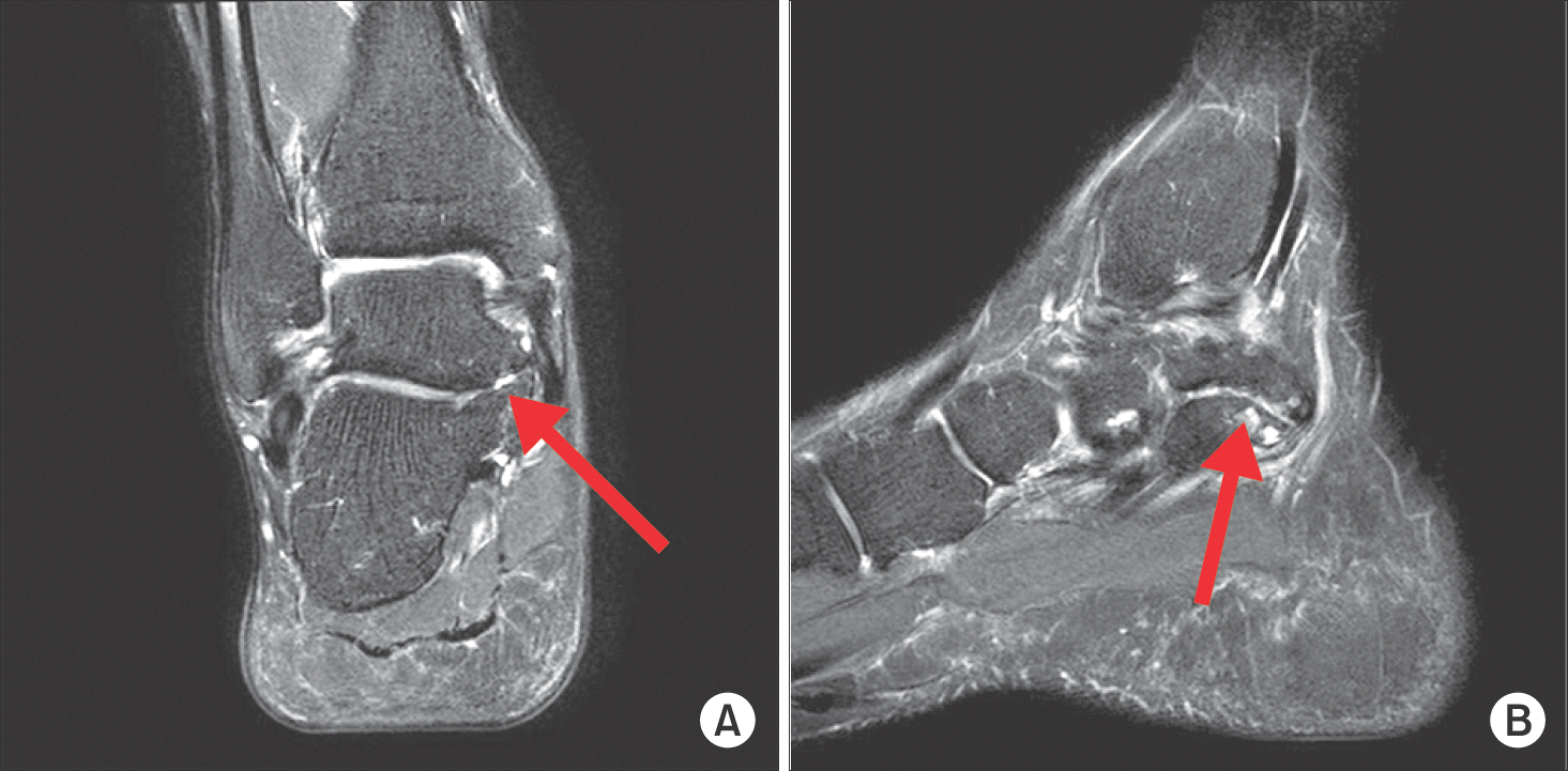 | Figure 1.Coronal (A) and sagittal (B) T2 image of the cartilagenous talocalcaneal coalition. In T2 images, there is high signal change in the border of the talus and calcaneus (arrows). |
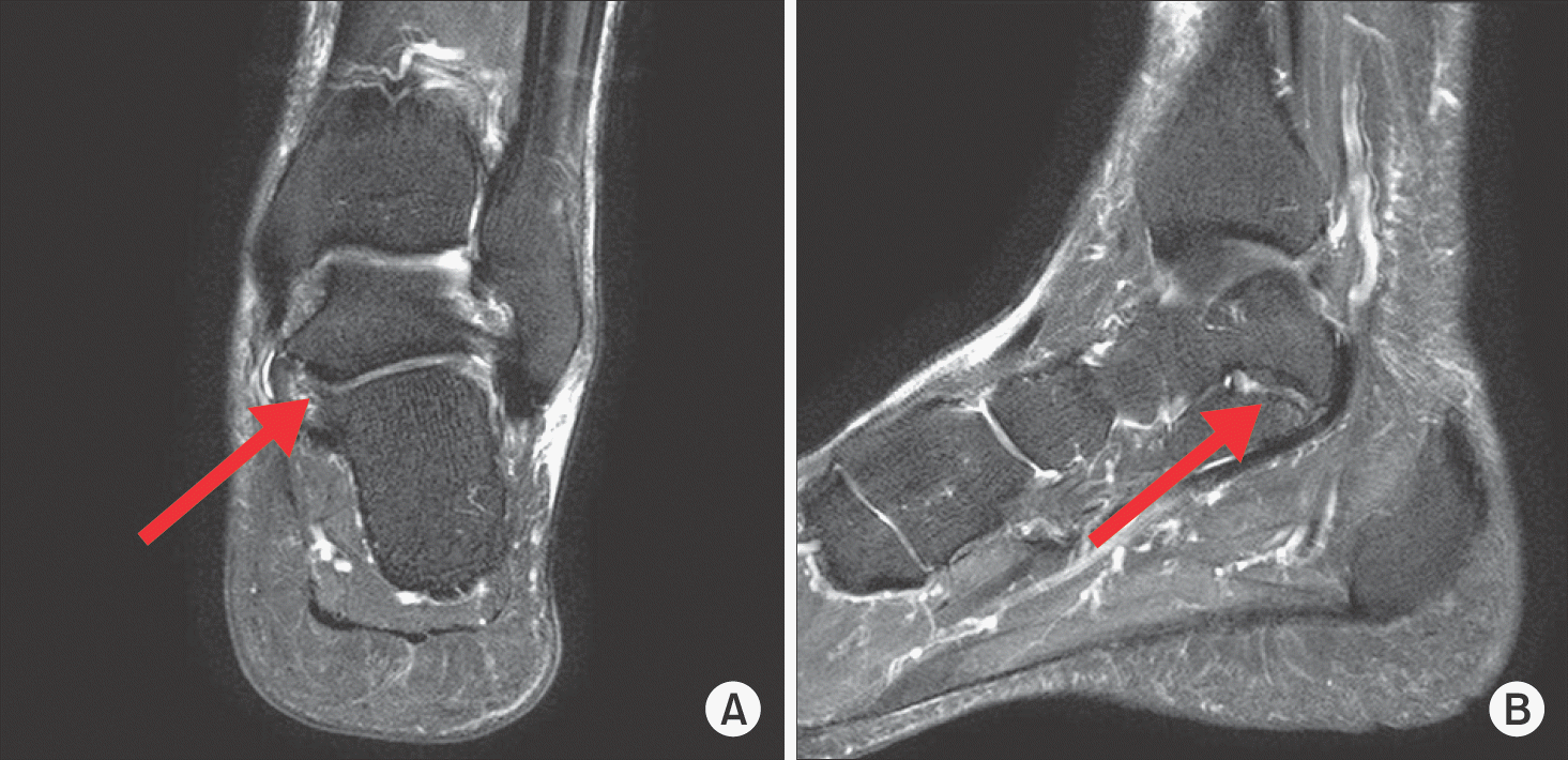 | Figure 2.Coronal (A) and sagittal (B) T2 image of the fibrous talonavicular coalition. In T2 images, there is low signal change in the border of the talus and calcaneus (arrows). |
 XML Download