Abstract
Purpose
We analyzed and compared the clinical and radiologic results between minimally invasive plate osteosynthesis and internal fixation using intramedullary (IM) nail in the treatment of distal tibia fractures.
Materials and Methods
From March 2005 to June 2013, 65 cases of distal tibia fractures treated with either plate fixation or IM nail fixation were analyzed retrospectively by clinical and radiologic evaluations. The clinical results were compared using the American Orthopaedic Foot and Ankle Society (AOFAS) score, Olerud-Molander ankle score (OMAS), and visual analogue scale (VAS) score at the last follow-up. The radiologic results were compared by time to bone union, complications such as nonunion, delayed union, and malunion.
Results
The clinical results (according to OMAS, AOFAS score, and VAS score) were 77.47, 84.76, and 1.75, respectively, in the plating group, and 90.21, 91.00, and 1.25, respectively, in the nailing group, and there was no statistically significant difference. Plating group showed earlier union than the nailing group and the nailing group showed higher frequency of non-union and delayed union than plating group.
Go to : 
References
1. Kim SK, Lee KB, Lim KY, Moon ES. Minimally invasive osteosynthesis with locking compression plate for distal tibia fractures. J Korean Fract Soc. 2011; 24:33–40.

2. Lee KB, Song SY, Kwon DJ, Lee YB, Rhee NK, Choi JH. A comparison between minimally invasive plate osteosynthesis & interlocking intramedullary nailing in distal tibia fractures. J Korean Fract Soc. 2008; 21:286–91.
3. Wyrsch B, McFerran MA, McAndrew M, Limbird TJ, Harper MC, Johnson KD, et al. Operative treatment of fractures of the tibial plafond. A randomized, prospective study. J Bone Joint Surg Am. 1996; 78:1646–57.

4. Chung ST, Kim HS, Cha SD, Yoo JH, Park JH, Kim JH, et al. Treatment of Distal tibia fracture using mippo technique with locking compression plate: comparative study of the intraarticular fracture and extraarticular fracture. J Korean Foot Ankle Soc. 2009; 13:162–8.
5. Edge AJ, Denham RA. External fixation for complicated tibial fractures. J Bone Joint Surg Br. 1981; 63-B:92–7.

6. Helfet DL, Shonnard PY, Levine D, Borrelli J Jr. Minimally invasive plate osteosynthesis of distal fractures of the tibia. Injury. 1997; 28(Suppl 1):A42–7. discussion A47–8.

7. Lau TW, Leung F, Chan CF, Chow SP. Wound complication of minimally invasive plate osteosynthesis in distal tibia fractures Int Orthop. 2008; 32:697–703.
8. Böstman O, Hänninen A. The fibular reciprocal fracture in tibial shaft fractures caused by indirect violence. Arch Orthop Trauma Surg. 1982; 100:115–21.
9. Konrath G, Moed BR, Watson JT, Kaneshiro S, Karges DE, Cramer KE. Intramedullary nailing of unstable diaphyseal fractures of the tibia with distal intraarticular involvement J Orthop Trauma. 1997; 11:200–5.
10. Tyllianakis M, Megas P, Giannikas D, Lambiris E. Interlocking intramedullary nailing in distal tibial fractures. Orthopedics. 2000; 23:805–8.

11. Nicoll EA. Closed and open management of tibial fractures. Clin Orthop Relat Res. 1974; 105:144–53.

12. Park IH, Song KW, Shin SI, Lee JY, Lee SY, Kim TH, et al. Interlocking intramedullary nailing for treating most distal tibial fracture J Korean Soc Fract. 2003; 16:356–62.
13. Robinson CM, McLauchlan GJ, McLean IP, Court-Brown CM. Distal metaphyseal fractures of the tibia with minimal involvement of the ankle1 Classification and treatment by locked intramedullary nailing. J Bone Joint Surg Br. 1995; 77:781–7.
14. Shon OJ, Chung SM. Interlocking intramedullary nail in distal tibia fracture. J Korean Fract Soc. 2007; 20:13–8.

15. Im GI, Tae SK. Distal metaphyseal fractures of tibia: a prospective randomized trial of closed reduction and intramedullary nail versus open reduction and plate and screws fixation. J Trauma. 2005; 59:1219–23. discussion 1223.

Go to : 
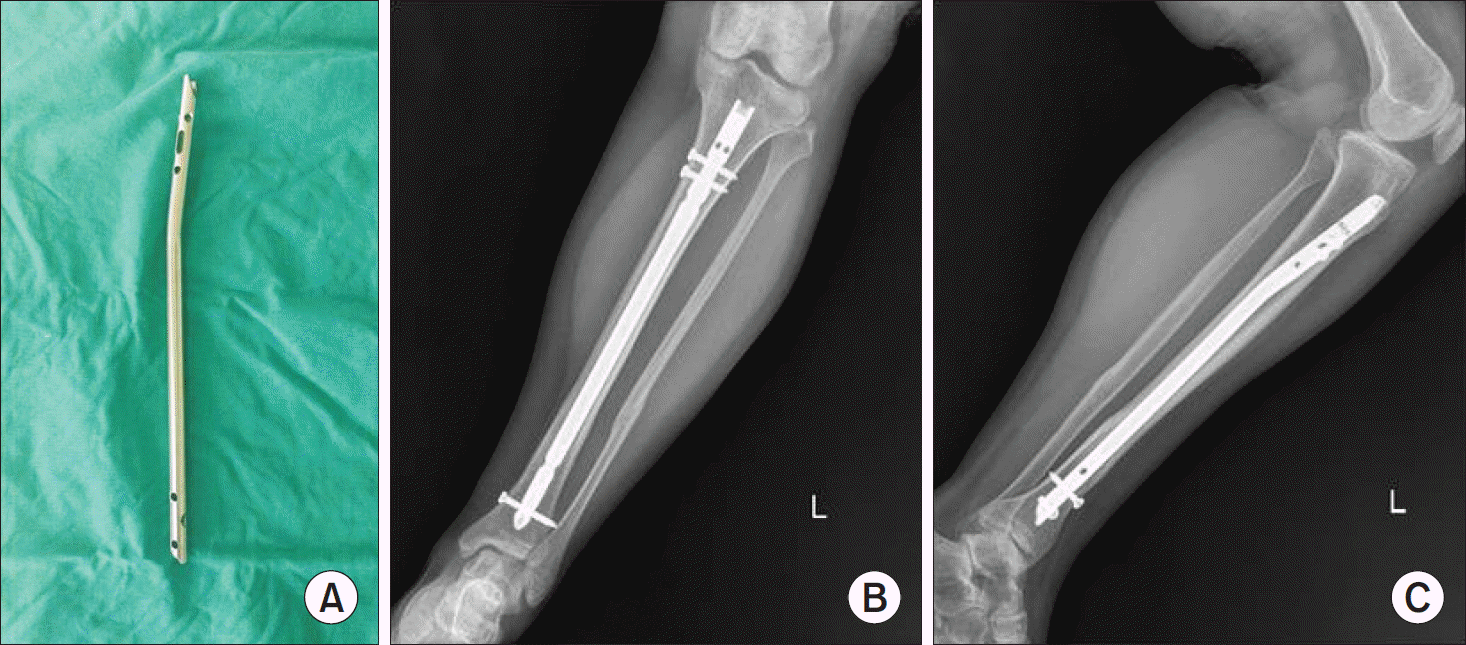
 | Figure 1.(A) This photograph demonstrates cannulated tibia nail (CTN; Synthes, Solothurn, Switzerland). (B) Anteroposterior view of the left tibia show stabilized distal tibial fracture by the CTN. (C) Lateral view of the left tibia show stabilized distal tibial fracture by the CTN. |
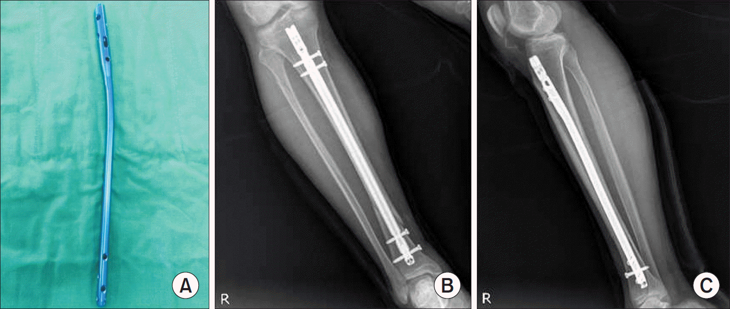
 | Figure 2.(A) This photograph demonstrates expert tibial nail (ETN; Synthes, Solothurn, Switzerland). (B) Anteroposterior view of the right tibia show stabilized distal tibial fracture by the ETN. (C) Lateral view of the right tibia show stabilized distal tibial fracture by the ETN. |
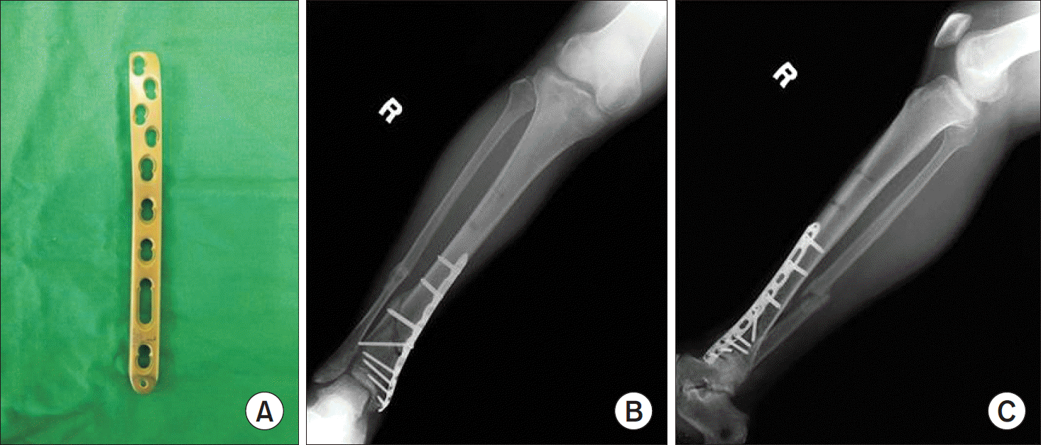
 | Figure 3.(A) This photograph demonstrate locking compression plate distal medial tibia (LCP-DMT; Synthes, Solo-thurn, Switzerland). (B) Anteroposterior view of the right tibia show stabilized distal tibial fracture by the LCP-DMT plate using minimal invasive technique. (C) Lateral view of the right tibia show stabilized distal tibial fracture by the LCP-DMT plate using minimal invasive technique. |
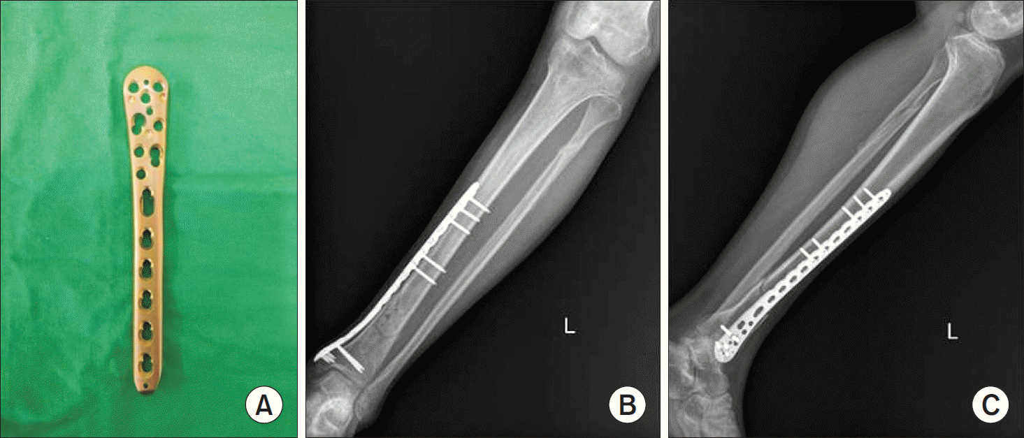
 | Figure 4.(A) This photograph demonstrate locking compression plate distal medial tibia low bend (LCP-DMT low bend; Synthes, Solothurn, Switzerland). (B) Anteroposterior view of the left tibia show stabilized distal tibial fracture by the LCP-DMT low bend plate using minimal invasive technique. (C) Lateral view of the left tibia show stabilized distal tibial fracture by the LCP-DMT low bend plate using minimal invasive technique. |
Table 1.
Demographics Data of Patients
| Plating group (n=25) | Nailing group (n=18) | |
|---|---|---|
| Mean age (yr) | 47 (20∼59) | 52 (24∼60) |
| Gender (male/female) | 14/11 | 10/8 |
| AO/OTA classification | ||
| 42-A1 | 2 | 6 |
| 42-A2 | 2 | 6 |
| 42-A3 | 6 | 5 |
| 42-C2 | 8 | 1 |
| 42-C3 | 7 | 0 |


 PDF
PDF ePub
ePub Citation
 Print
Print


 XML Download