

 PDF
PDF ePub
ePub Citation
 Print
Print


Abstract
Purpose:
The purpose of this study was to report impacts of the amount of displacement of percutaneous osteotomy on the clinical and radiologic results in the treatment of bunionette deformities.
Materials and Methods:
We retrospectively reviewed 36 cases of bunionette deformities treated with percutaneous modified Kramer osteotomies from 2009 to 2013. We measured amounts of displacement on anteroposterior and lateral plain radiographs as well as multiple parameters which represent degrees of the bunionette deformities. We also recorded radiological healing time, clinical healing time, residual symptoms, and the time of returning to daily activity.
Results:
No meaningful correlation was found between severity of preoperative deformity and amount of displacement of the osteotomy. The amount of displacement on a horizontal plane did not affect the healing time, duration of symptoms, or time of returning to daily activity. However, large sagittal displacement was related to duration of postoperative symptoms.
Conclusion:
Findings of this study suggest that the displacement in percutaneous osteotomy for bunionette deformity does not affect clinical results and healing time. We believe that we do not need to be excessively cautious about how large the displacement we make during the percutaneous modified Kramer osteotomy for the bunionette deformity.
Go to : 
REFERENCES
1.Ajis A., Koti M., Maffulli N. Tailor’s bunion: a review. J Foot Ankle Surg. 2005. 44:236–45.

2.Cohen BE., Nicholson CW. Bunionette deformity. J Am Acad Or-thop Surg. 2007. 15:300–7.
3.Kim YC., Ahn JH. Bunionette deformity. J Korean Foot Ankle Soc. 2013. 17:1–10.
4.Mann RA., Mann JA. The bunionette deformity. Instr Course Lect. 2004. 53:303–9.
5.Coughlin MJ. Treatment of bunionette deformity with longitudinal diaphyseal osteotomy with distal soft tissue repair. Foot Ankle. 1991. 11:195–203.
6.Kitaoka HB., Holiday AD Jr. Metatarsal head resection for bunionette: long-term follow-up. Foot Ankle. 1991. 11:345–9.
7.Cooper MT., Coughlin MJ. Subcaptial oblique fifth metatarsal osteotomy versus distal chevron osteotomy for correction of bunionette deformity: a cadaveric study. Foot Ankle Spec. 2012. 5:313–7.
8.Lee KT., Young KW., Kim JY., Cha SD., Kim ES., Ahn YJ. Treatment of bunionette deformity with distal chevron osteotomy. J Korean Orthop Assoc. 2006. 41:14–8.
9.Glover JP., Weil L Jr., Weil LS Sr. Scarfette osteotomy for surgical treatment of bunionette deformity. Foot Ankle Spec. 2009. 2:73–8.
10.Guha AR., Mukhopadhyay S., Thomas RH. ‘Reverse’ scarf osteotomy for bunionette correction: initial results of a new surgical technique. Foot Ankle Surg. 2012. 18:50–4.
11.Seide HW., Petersen W. Tailor’s bunion: results of a scarf osteotomy for the correction of an increased intermetatarsal IV/V angle. A report on ten cases with a 1-year follow-up. Arch Or-thop Trauma Surg. 2001. 121:166–9.
12.Ahn JH., Kim HY., Kang JW., Choy WS., Kim YI. Treatment of bunionette deformity with diaphyseal oblique osteotomy. J Korean Foot Ankle Soc. 2008. 12:31–5.
13.Kim SK., Kim J., Lee JI., Rhee SH. Treatment of bunionette deformity with diaphyseal oblique osteotomy. J Korean Foot Ankle Soc. 2014. 18:19–23.
14.London BP., Stern SF., Quist MA., Lee RK., Picklesimer EK. Long oblique distal osteotomy of the fifth metatarsal for correction of tailor’s bunion: a retrospective review. J Foot Ankle Surg. 2003. 42:36–42.
15.Okuda R., Kinoshita M., Morikawa J., Jotoku T., Abe M. Proximal dome-shaped osteotomy for symptomatic bunionette. Clin Or-thop Relat Res. 2002. 396:173–8.
16.Baumhauer JF., DiGiovanni BF. Osteotomies of the fifth metatarsal. Foot Ankle Clin. 2001. 6:491–8.
17.Haddon TB., LaPointe SJ. Relative strength of tailor’s bunion osteotomies and fixation techniques. J Foot Ankle Surg. 2013. 52:16–23.
18.Weil L Jr, Weil LS Sr. Osteotomies for bunionette deformity. Foot Ankle Clin. 2011. 16:689–712.
19.Waizy H., Olender G., Mansouri F., Floerkemeier T., Stukenborg-Colsman C. Minimally invasive osteotomy for symptomatic bunionette deformity is not advisable for severe deformities: a critical retrospective analysis of the results. Foot Ankle Spec. 2012. 5:91–6.
20.Kim SY., Park KH., Lee JW. Treatment of bunionette deformity with S.E.R.I. (simple, effective, rapid, inexpensive) operation. J Korean Foot Ankle Soc. 2010. 14:25–30.
21.Giannini S., Faldini C., Nanni M., Di Martino A., Luciani D., Vannini F. A minimally invasive technique for surgical treatment of hallux valgus: simple, effective, rapid, inexpensive (SERI). Int Orthop. 2013. 37:1805–13.
22.Laffenêtre O., Millet-Barbé B., Darcel V., Lucas Y Hernandez J., Chauveaux D. Percutaneous bunionette correction: results of a 49-case retrospective study at a mean 34 months’ follow-up. Orthop Traumatol Surg Res. 2015. 101:179–84.
23.Lui TH. Percutaneous osteotomy of the fifth metatarsal for symptomatic bunionette. J Foot Ankle Surg. 2014. 53:747–52.
24.Magnan B., Samaila E., Merlini M., Bondi M., Mezzari S., Bartolozzi P. Percutaneous distal osteotomy of the fifth metatarsal for correction of bunionette. J Bone Joint Surg Am. 2011. 93:2116–22.
25.Michels F., Van Der Bauwhede J., Guillo S., Oosterlinck D., de Lavigne C. Percutaneous bunionette correction. Foot Ankle Surg. 2013. 19:9–14.
26.Magnan B., Bondi M., Mezzari S., Bonetti I., Samaila E. Minimally invasive surgery of the forefoot: current concept review. Int J Clin Med. 2013. 4:11–9.
27.Legenstein R., Bonomo J., Huber W., Boesch P. Correction of tailor’s bunion with the Boesch technique: a retrospective study. Foot Ankle Int. 2007. 28:799–803.
28.Weitzel S., Trnka HJ., Petroutsas J. Transverse medial slide osteotomy for bunionette deformity: long-term results. Foot Ankle Int. 2007. 28:794–8.
Go to :
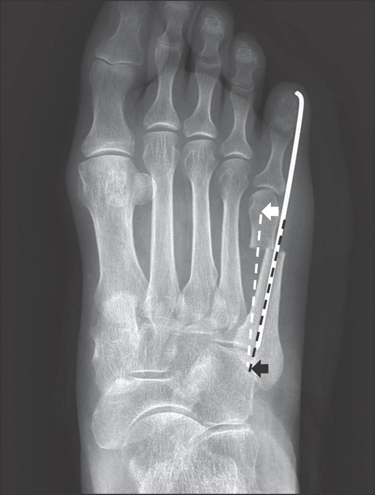 | Figure 1.Metatarsal angle line was defined by the line (white dotted line) from the center of the metatarsal head (white arrow) to the point (black arrow) on which proximal diaphyseal dissecting line (black dotted line) cross the proximal joint. |
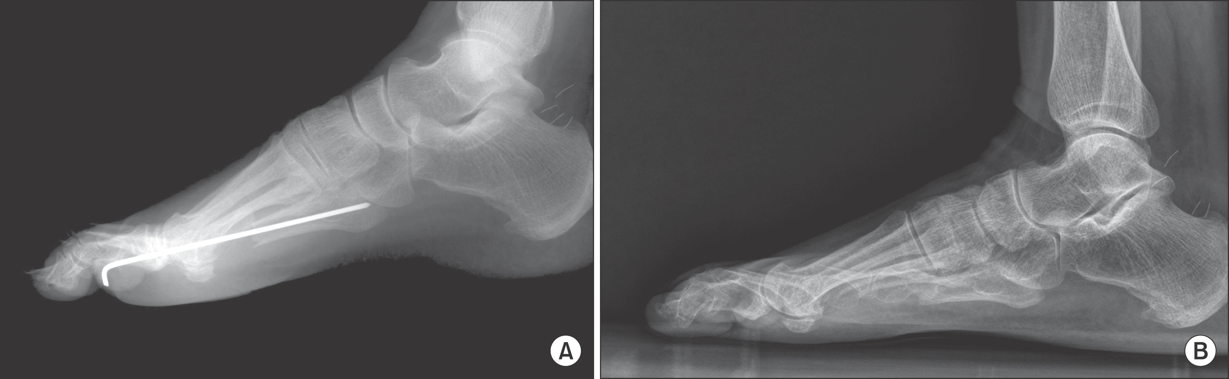 | Figure 2.In this case we made big sagittal displacement during the procedure (A) and vague pain was persisted until 16 weeks after the surgery (B). |
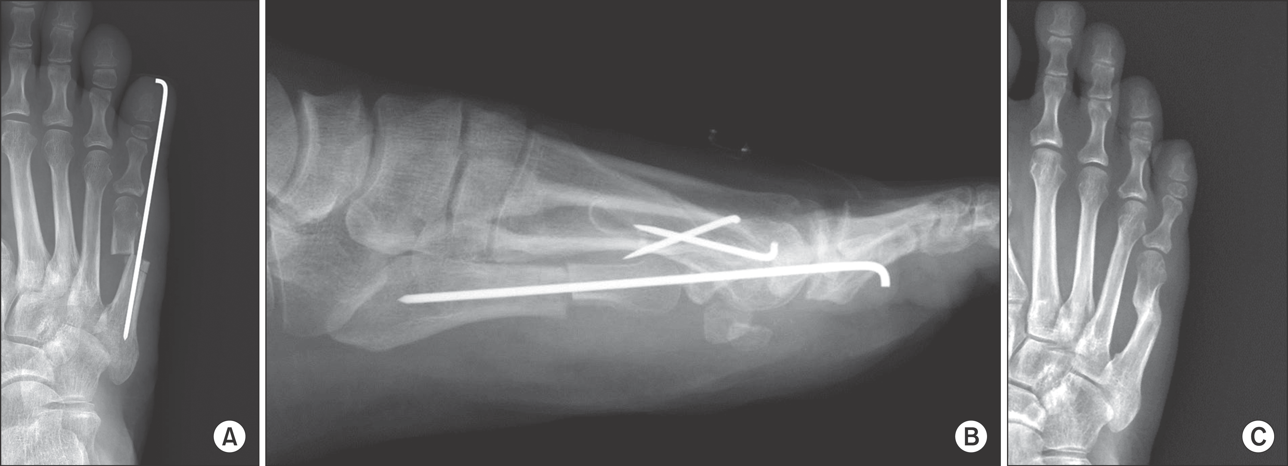 | Figure 3.In this case we made huge amount of displacement of metatarsal osteotomy in horizontal plane (A) but not in sagittal plane (B). Follow-up radiograph shows that such a big displacement did not affect bone healing (C). |
Table 1.
Amount of Displacement in Each Fallat Type of Bunionette Deformity
| Fallat type | Mean value of displacement (%) | |
|---|---|---|
| Horizontal | Sagittal | |
| I | 57.2 | 31.9 |
| II | 54.6 | 31.2 |
| III | 54.8 | 30.4 |
| IV | 56.6 | 31.5 |
Table 2.
Parameters Represent Degrees of Bunionette Deformity
| Parameter | Preoperative | Postoperative | Last follow-up |
|---|---|---|---|
| 2nd∼5th IMA (°) | 20.48±0.89 | 8.75±1.46 | 12.93±0.74 |
| 4th∼5th IMA (°) | 10.85±0.47 | 3.53±0.43 | 4.68±0.44 |
| 5th MTPA (°) | 14.92±0.89 | 10.16±0.76 | 4.55±0.60 |
Table 3.
Correlation between Amount of Displacement in Each Plane and Degrees of Deformity
Table 4.
Correlation between Amount of Displacement in Each Plane and Clinical Parameters
| Plane of displacement | Radiologic healing time | Duration of symptom | Time of returning to normal activity | |||
|---|---|---|---|---|---|---|
| Pearson coefficient | p-value | Pearson coefficient | p-value | Pearson coefficient | p-value | |
| Horizontal | -0.223 | 0.192 | -0.258 | 0.128 | 0.033 | 0.850 |
| Sagittal | 0.180 | 0.293 | 0.387 | 0.020* | 0.224 | 0.189 |
Table 5.
Comparing Parameters between Delayed Union Group and Normal Union Group by Independent t-test
| Parameter | Delayed union group | Normal union group | p-value |
|---|---|---|---|
| Dispacement in horizontal plane (%) | 49.0±26.0 | 60.0±14.4 | 0.283 |
| Displacement in sagittal plane (%) | 35.4±31.4 | 23.0±20.0 | 0.185 |
| Duration of symptom (wk) | 8.2±3.8 | 4.5±1.9 | 0.031* |
| Time of returning to normal activity (wk) | 6.9±1.5 | 5.7±1.3 | 0.030 * |
 XML Download