

 PDF
PDF ePub
ePub Citation
 Print
Print


Abstract
Purpose:
The purpose of this report is to investigate the clinical and radiological results of corrective osteotomy of the 3rd metatarsal bone for shortening and dorsal displacement without exposure around neuroma.
Materials and Methods:
Twelve cases of patients who underwent corrective osteotomy of metatarsal bone for a Morton’s neuroma from November 2013 to September 2014 were retrospectively reviewed. Corrective osteotomy was performed through a dorsal approach at the 3rd metatarsal bone base and distal metatarsal bone was displaced dorsally and proximally. Preoperative and postoperative pain assessed using American Orthopaedic Foot and Ankle Society (AOFAS) score and radiographs were evaluated.
REFERENCES
1.Gauthier G. Thomas Morton’s disease: a nerve entrapment syndrome. A new surgical technique. Clin Orthop Relat Res. 1979. 142:90–2.
2.Rasmussen MR., Kitaoka HB., Patzer GL. Nonoperative treatment of plantar interdigital neuroma with a single corticosteroid injection. Clin Orthop Relat Res. 1996. 326:188–93.

3.Kay D., Bennett GL. Morton’s neuroma. Foot Ankle Clin. 2003. 8:49–59.
4.Bennett GL., Graham CE., Mauldin DM. Morton’s interdigital neuroma: a comprehensive treatment protocol. Foot Ankle Int. 1995. 16:760–3.
5.Shapiro SL. Endoscopic decompression of the intermetatarsal nerve for Morton’s neuroma. Foot Ankle Clin. 2004. 9:297–304.
6.Kasparek M., Schneider W. Surgical treatment of Morton’s neuroma: clinical results after open excision. Int Orthop. 2013. 37:1857–61.
7.Fabié F., Accadbled F., Tricoire JL., Puget J. Anatomic danger of percutaneous section of the inter-metatarsal ligament for the treatment of Morton’s neuroma. Rev Chir Orthop Reparatrice Appar Mot. 2007. 93:720–4.
8.Zelent ME., Kane RM., Neese DJ., Lockner WB. Minimally invasive Morton’s intermetatarsal neuroma decompression. Foot Ankle Int. 2007. 28:263–5.
9.Stamatis ED., Karabalis C. Interdigital neuromas: current state of the art--surgical. Foot Ankle Clin. 2004. 9:287–96.
10.Maestro M., Besse JL., Ragusa M., Berthonnaud E. Forefoot mor-photype study and planning method for forefoot osteotomy. Foot Ankle Clin. 2003. 8:695–710.
11.Mulder JD. The causative mechanism in morton’s metatarsalgia. J Bone Joint Surg Br. 1951. 33-B:94–5.
12.Mendicino SS., Rockett MS. Morton’s neuroma. Update on diagnosis and imaging. Clin Podiatr Med Surg. 1997. 14:303–11.
13.Sharp RJ., Wade CM., Hennessy MS., Saxby TS. The role of MRI and ultrasound imaging in Morton’s neuroma and the effect of size of lesion on symptoms. J Bone Joint Surg Br. 2003. 85:999–1005.
14.Coughlin MJ., Pinsonneault T. Operative treatment of interdigital neuroma. A long-term follow-up study. J Bone Joint Surg Am. 2001. 83:1321–8.
15.Johnson JE., Johnson KA., Unni KK. Persistent pain after excision of an interdigital neuroma. Results of reoperation. J Bone Joint Surg Am. 1988. 70:651–7.
16.Akermark C., Saartok T., Zuber Z. A prospective 2-year followup study of plantar incisions in the treatment of primary intermetatarsal neuromas (Morton’s neuroma). Foot Ankle Surg. 2008. 14:67–73.
17.Kim JY., Choi JH., Park J., Wang J., Lee I. An anatomical study of Morton’s interdigital neuroma: the relationship between the occurring site and the deep transverse metatarsal ligament (DTML). Foot Ankle Int. 2007. 28:1007–10.
18.Hofstaetter SG., Hofstaetter JG., Petroutsas JA., Gruber F., Ritschl P., Trnka HJ. The Weil osteotomy: a seven-year follow-up. J Bone Joint Surg Br. 2005. 87:1507–11.
19.Khurana A., Kadamabande S., James S., Tanaka H., Hariharan K. Weil osteotomy: assessment of medium term results and predictive factors in recurrent metatarsalgia. Foot Ankle Surg. 2011. 17:150–7.
20.Bauer T., Gaumetou E., Klouche S., Hardy P., Maffulli N. Metatarsalgia and Morton’s disease: comparison of outcomes between open procedure and neurectomy versus percutaneous metatarsal osteotomies and ligament release with a minimum of 2 years of follow-up. J Foot Ankle Surg. 2015. 54:373–7.
21.Myerson MS. The diagnosis and treatment of injury to the tarsometatarsal joint complex. J Bone Joint Surg Br. 1999. 81:756–63.
Figure 2.
Method for the measurement of 3rd metatarsal length. Third metatarsal length (dashed line) was determined by measuring the distances from apex of each metatarsal to transmetatarsal line centered from lateral sesamoid and perpendicular to the second metatarsal axis.
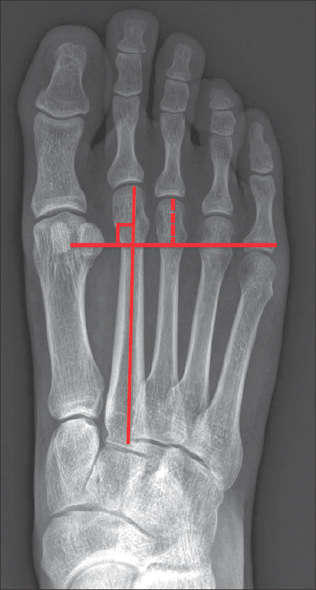
Figure 5.
C-arm radiographs. (A) Preoperative sesamoid view. (B) Postoperative sesamoid view (arrow). Third metatarsal bone was dorsally displaced after the metatarsal corrective osteotomy.
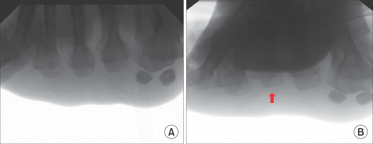
Figure 6.
(A) Preoperative foot standing anteroposterior (AP) radiograph. (B) Postoperative foot standing AP radiograph. After the metatarsal corrective osteotomy, the length of 3rd metatarsal bone was shortened.
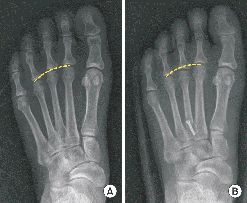
Table 1.
Summary of Cases
Table 2.
Functional and Radiologic Results
 XML Download