

 PDF
PDF ePub
ePub Citation
 Print
Print


Abstract
Purpose:
Minimal incision distal metatarsal osteotomy (MIDMO) is known to be an effective surgical procedure for mild to moderate hallux valgus. However, the result of MIDMO on moderate to severe hallux valgus is controversial; therefore, we investigated the radiological and clinical results of MIDMO on moderate to severe hallux valgus.
Materials and Methods:
We reviewed 51 feet (48 patients) with moderate to severe hallux valgus. The mean age was 67.0 years and the mean follow-up period was 32.2 months. Radiological data of hallux valgus angle, first intermetatarsal angle, and distal metatarsal articular angle on plain radiographs were analyzed. Recurrence, union, lateral translation of distal fragment and angulation were also analyzed. The clinical data were obtained using American Orthopaedic Foot and Ankle Society (AOFAS) score of preoperation and last follow-up. Receiver operating characteristic (ROC) curve was used to determine a cut-off value.
Results:
The mean hallux valgus angle measured at preoperation was 37.7° and 15.9° at last follow-up. The mean first intermetatarsal angle of preoperation and last follow-up were 15.2° and 8.3°. The mean distal metatarsal articular angle changed from 12.6° at preoperation to 7.8° at last follow-up. Preoperative hallux valgus angle (p=0.0051) and distal metatarsal articular angle (p=0.0078) were statistically significant factors affecting postoperative AOFAS score. Cut-off value of each was 37° and 13°, respectively. Lateral translation of distal fragment in 5 recurrent cases was 23.0% compared to 45.3% of 46 non-recurrent cases. The result was statistically significant and the cut-off value was 38%.
REFERENCES
1.Robinson AH., Limbers JP. Modern concepts in the treatment of hallux valgus. J Bone Joint Surg Br. 2005. 87:1038–45.

2.Radwan YA., Mansour AM. Percutaneous distal metatarsal osteotomy versus distal chevron osteotomy for correction of mild-to-moderate hallux valgus deformity. Arch Orthop Trauma Surg. 2012. 132:1539–46.
3.Borton DC., Stephens MM. Basal metatarsal osteotomy for hallux valgus. J Bone Joint Surg Br. 1994. 76:204–9.
4.Coughlin MJ., Grebing BR., Jones CP. Arthrodesis of the first metatarsophalangeal joint for idiopathic hallux valgus: intermediate results. Foot Ankle Int. 2005. 26:783–92.
5.Klosok JK., Pring DJ., Jessop JH., Maffulli N. Chevron or Wilson metatarsal osteotomy for hallux valgus. A prospective randomised trial. J Bone Joint Surg Br. 1993. 75:825–9.
6.Sammarco GJ., Brainard BJ., Sammarco VJ. Bunion correction using proximal Chevron osteotomy. Foot Ankle. 1993. 14:8–14.
7.Bösch P., Markowski H., Rannicher V. Technik und erste ergebnisse der subkutanen distalen metatarsale-I-osteotomie. Orthop Praxis. 1990. 26:51–6.
8.Faour-Martín O., Martín-Ferrero MA., Valverde García JA., Vega-Castrillo A., de la Red-Gallego MA. Long-term results of the ret-rocapital metatarsal percutaneous osteotomy for hallux valgus. Int Orthop. 2013. 37:1799–803.
9.Bösch P., Wanke S., Legenstein R. Hallux valgus correction by the method of Bösch: a new technique with a seven-to-ten-year follow-up. Foot Ankle Clin. 2000. 5:485–98.
10.Kissel CG., Unroe BJ., Parker RM. The offset “V” bunionectomy using cortical screw and buried Kirschner wire fixation. J Foot Surg. 1992. 31:560–77.
11.Magnan B., Pezzè L., Rossi N., Bartolozzi P. Percutaneous distal metatarsal osteotomy for correction of hallux valgus. J Bone Joint Surg Am. 2005. 87:1191–9.
12.Kadakia AR., Smerek JP., Myerson MS. Radiographic results after percutaneous distal metatarsal osteotomy for correction of hallux valgus deformity. Foot Ankle Int. 2007. 28:355–60.
13.Huang PJ., Lin YC., Fu YC., Yang YH., Cheng YM. Radiographic evaluation of minimally invasive distal metatarsal osteotomy for hallux valgus. Foot Ankle Int. 2011. 32:S503–7.
14.Angthong C., Yoshimura I., Kanazawa K., Hagio T., Ida T., Naito M. Minimally invasive distal linear metatarsal osteotomy for correction of hallux valgus: a preliminary study of clinical outcome and analytical radiographic results via a mapping system. Arch Orthop Trauma Surg. 2013. 133:321–31.
15.Ozan F., Bora OA., Filiz MA., Kement Z. Interposition arthroplasty in the treatment of hallux rigidus. Acta Orthop Traumatol Turc. 2010. 44:143–51.
16.Hawkins FB., Mitchell CL., Hedrick DW. Correction of hallux valgus by metatarsal osteotomy. J Bone Joint Surg Am. 1945. 27:387–94.
17.Mann RA., Coughlin MJ. Hallux valgus--etiology, anatomy, treatment and surgical considerations. Clin Orthop Relat Res. 1981. 157:31–41.
18.Shapiro F., Heller L. The Mitchell distal metatarsal osteotomy in the treatment of hallux valgus. Clin Orthop Relat Res. 1975. 107:225–31.
19.Trnka HJ., Zembsch A., Easley ME., Salzer M., Ritschl P., Myerson MS. The chevron osteotomy for correction of hallux valgus. Comparison of findings after two and five years of follow-up. J Bone Joint Surg Am. 2000. 82:1373–8.
20.Wu KK. Wu’s bunionectomy: a clinical analysis of 150 personal cases. J Foot Surg. 1992. 31:288–97.
21.Giannini S., Faldini C., Nanni M., Di Martino A., Luciani D., Vannini F. A minimally invasive technique for surgical treatment of hallux valgus: simple, effective, rapid, inexpensive (SERI). Int Orthop. 2013. 37:1805–13.
22.Portaluri M. Hallux valgus correction by the method of Bösch: a clinical evaluation. Foot Ankle Clin. 2000. 5:499–511.
23.Mann RA., Donatto KC. The chevron osteotomy: a clinical and radiographic analysis. Foot Ankle Int. 1997. 18:255–61.
24.Sanhudo JA. Extending the indications for distal chevron osteotomy. Foot Ankle Int. 2000. 21:522–3.
25.Bonnel F., Canovas F., Poirée G., Dusserre F., Vergnes C. Evaluation of the Scarf osteotomy in hallux valgus related to distal metatarsal articular angle: a prospective study of 79 operated cases. Rev Chir Orthop Reparatrice Appar Mot. 1999. 85:381–6.
26.Pontious J., Mahan KT., Carter S. Characteristics of adolescent hallux abducto valgus. A retrospective review. J Am Podiatr Med Assoc. 1994. 84:208–18.
27.Okuda R., Kinoshita M., Yasuda T., Jotoku T., Kitano N., Shima H. Postoperative incomplete reduction of the sesamoids as a risk factor for recurrence of hallux valgus. J Bone Joint Surg Am. 2009. 91:1637–45.
28.Veri JP., Pirani SP., Claridge R. Crescentic proximal metatarsal osteotomy for moderate to severe hallux valgus: a mean 12.2 year follow-up study. Foot Ankle Int. 2001. 22:817–22.
29.Deenik AR., de Visser E., Louwerens JW., de Waal Malefijt M., Drai-jer FF., de Bie RA. Hallux valgus angle as main predictor for correction of hallux valgus. BMC Musculoskelet Disord. 2008. 9:70.
Figure 1.
The lines are osteotomy site and skin incision line of minimal incision distal metatarsal osteotomy. M-P joint: metatarsophalangeal joint.
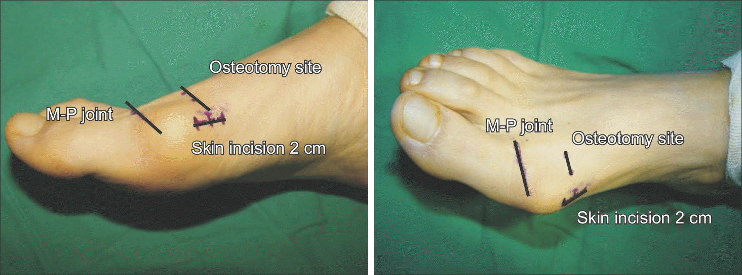
Figure 2.
After inserting Kirschner wire from the incision site into the soft tissue of hallus in proximal-todistal direction along the axis, insert the Kirschner wire in the opposite direction.
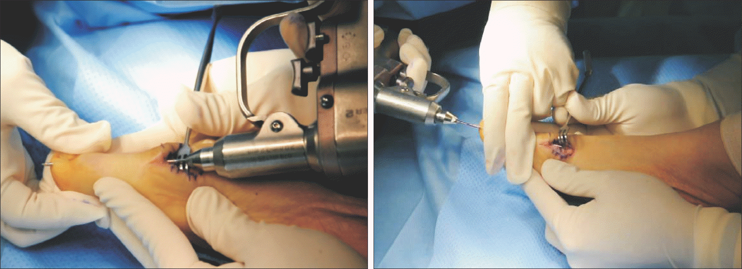
Figure 3.
The left foot of 59-year-old female was treated with minimal incision distal metatarsal osteotomy. Anteroposterior radiograph of preoperative (A), postoperative (B), and last follow-up (C).
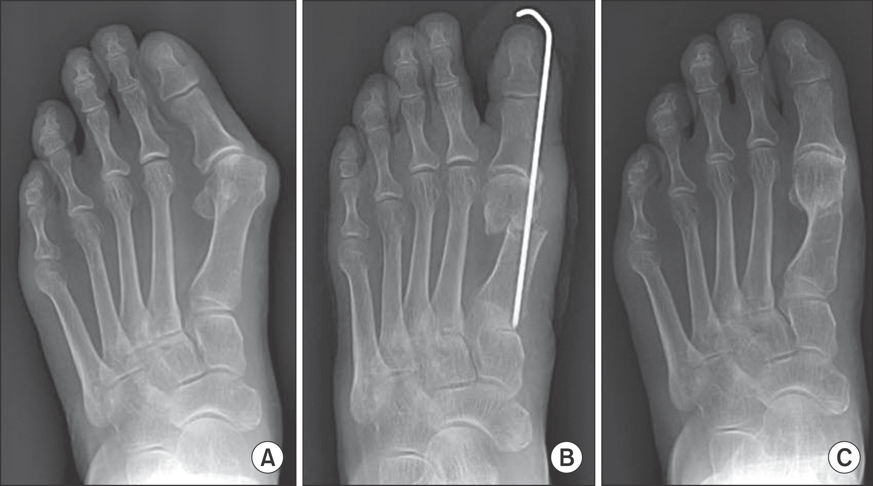
Figure 4.
Receiver operating characteristic curve of recurrence and lateral translation is shown that 38% is proposed as cut-off value. CI: confidence interval, +LR: positive likelihood ratio.
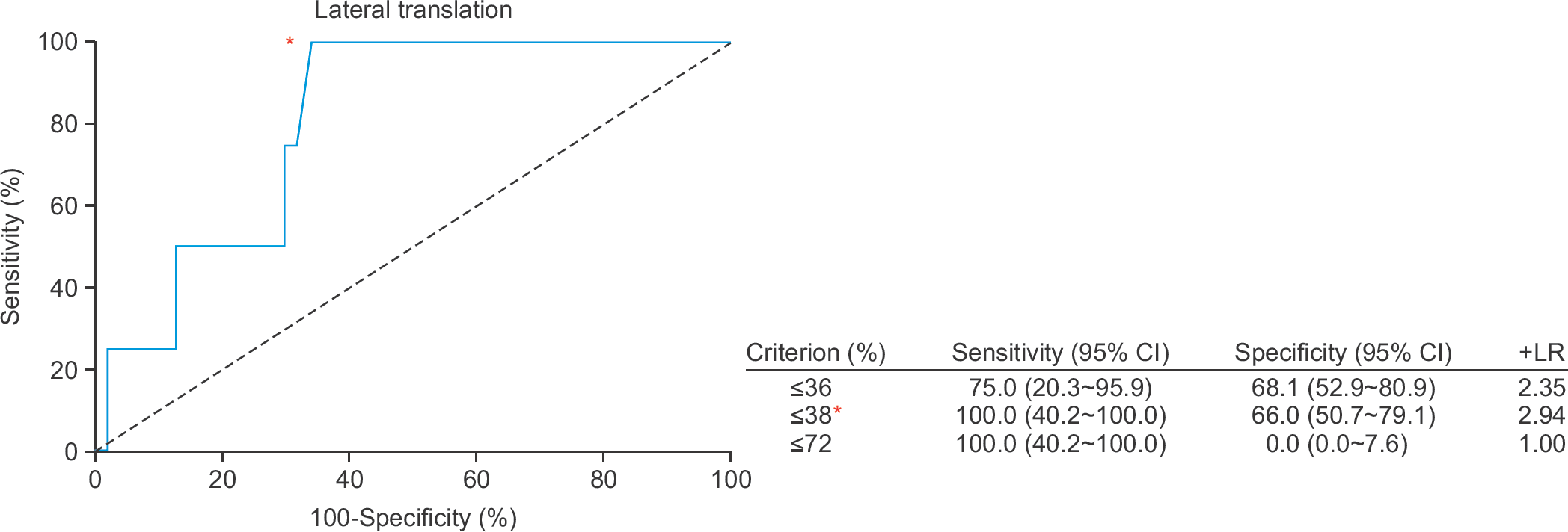
Figure 5.
Receiver operating characteristic curve of American Orthopaedic Foot and Ankle Society score and preoperative hallux valgus angle (HVA) is shown that 37° is proposed as cut-off value. CI: confidence interval, +LR: positive likelihood ratio.
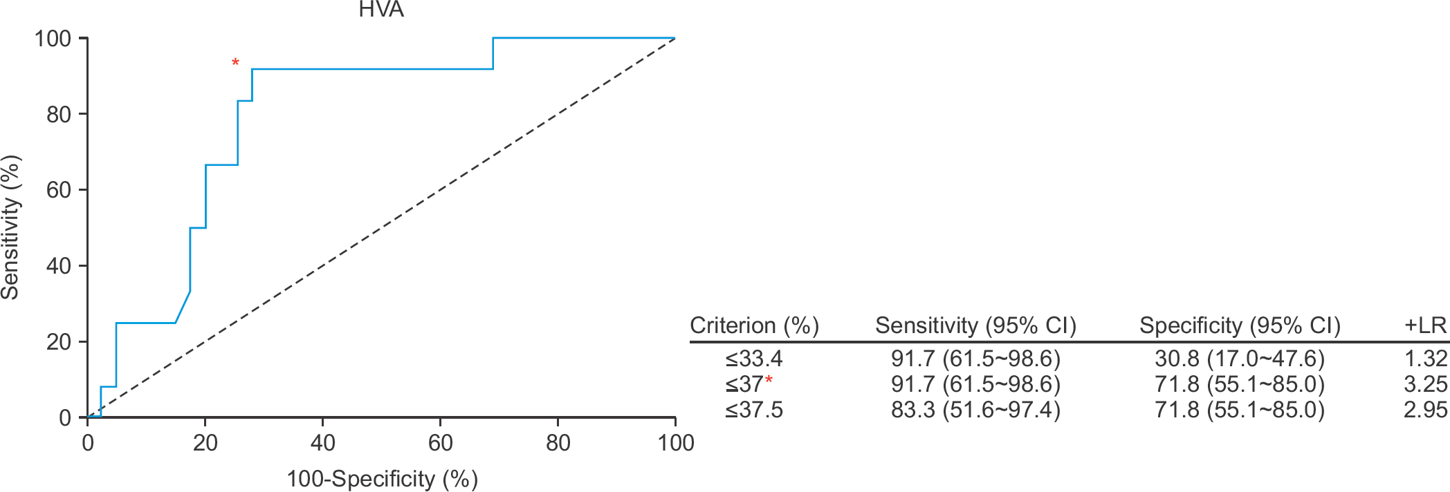
Figure 6.
Receiver operating characteristic curve of American Orthopaedic Foot and Ankle Society score and preoperative distal metatarsal articular angle (DMAA) is shown that 13° is proposed as cut-off value. CI: confidence interval, +LR: positive likelihood ratio.
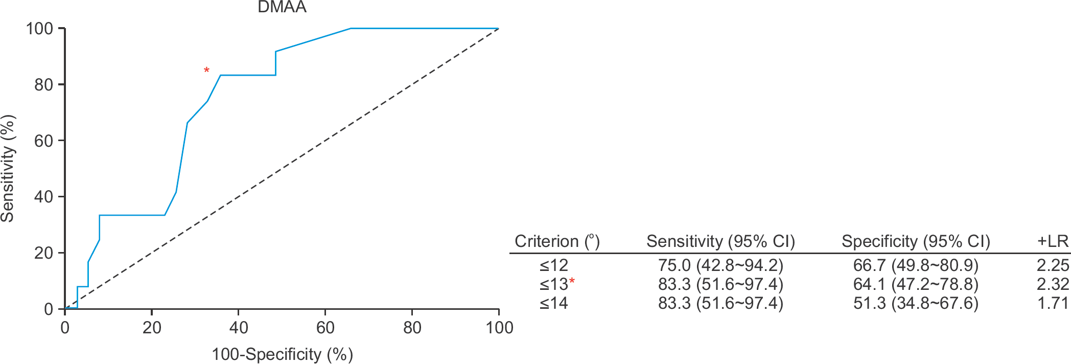
Table 1.
Radiologic Data after Minimal Incision Distal Metatarsal Osteotomy
| Angle (°) | Preoperative | Postoperative | After pin removal | Follow-up | Last follow-up | ||
|---|---|---|---|---|---|---|---|
| 3 mo | 6 mo | 1 yr | |||||
| HVA | 37.7 | ―2.8 | 8.6 | 12.8 | 14.7 | 15.5 | 15.9 |
| IMA | 15.2 | 4.2 | 5.8 | 6.0 | 6.3 | 6.4 | 8.3 |
| DMAA | 12.6 | 1.1 | 4.1 | 6.6 | 7.9 | 8.5 | 7.8 |
Table 2.
Comparison of Recurrence and Non-recurrence Group
| Lateral translation (%) | Mean preoperative HVA (°) | |
|---|---|---|
| Recurrence (n=5) | 23.0 | 39.4 |
| Non-recurrence (n=46) | 45.3 | 35.6 |
| p-value* | 0.0045 | 0.1046 |
Table 3.
Factors Affecting and Not Affecting AOFAS Score in Minima Incision Distal Metatarsal Osteotomy
 XML Download