Abstract
Purpose
Tibiotalocalcaneal arthrodesis has been used as a treatment option for severe deformity including Charcot arthropathy, avascular necrosis of the talus, and severe osteoarthritis of the ankle and subtalar joint. The purpose of this study was to evaluate the result of the surgical outcome of tibiotalocalcaneal arthrodesis using a retrograde intramedullary nail.
Materials and Methods
Tibiotalocalcaneal arthrodesis using a retrograde intramedullary nail was performed by one surgeon in 36 cases. Clinical and radiological finding was evaluated using assessment of fusion time, 5th metatarsal-tibial angle, possibility of postoperative complication, visual analogue scale for pain and American Orthopaedic Foot and Ankle Society (AOFAS) score.
Results
Union was achieved in 33 cases at an average of 23 weeks (11∼29 weeks). There were 3 cases of nonunion and 1 case of reoperation. Nail-tibial angle tended to be larger in nonunion cases. AOFAS score showed significantly poor outcome at malalignment (≥5o), negative value of 5th metatarsal-tibial angle.
References
1. Pelton K, Hofer JK, Thordarson DB. Tibiotalocalcaneal arthrodesis using a dynamically locked retrograde intramedullary nail. Foot Ankle Int. 2006; 27:759–63.

3. Alfahd U, Roth SE, Stephen D, Whyne CM. Biomechanical comparison of intramedullary nail and blade plate fixation for tibiotalocalcaneal arthrodesis. J Orthop Trauma. 2005; 19:703–8.

4. Chiodo CP, Acevedo JI, Sammarco VJ, Parks BG, Boucher HR, Myerson MS, et al. Intramedullary rod fixation compared with blade-plate-and-screw fixation for tibiotalocalcaneal arthrodesis: a biomechanical investigation. J Bone Joint Surg Am. 2003; 85:2425–8.
5. Berend ME, Glisson RR, Nunley JA. A biomechanical comparison of intramedullary nail and crossed lag screw fixation for tibiotalocalcaneal arthrodesis. Foot Ankle Int. 1997; 18:639–43.

6. Gerstenfeld LC, Cullinane DM, Barnes GL, Graves DT, Einhorn TA. Fracture healing as a postnatal developmental process: molecular, spatial, and temporal aspects of its regulation. J Cell Biochem1. 2003; 88:873–84.

7. Mann RA, Rongstad KM. Arthrodesis of the ankle: a critical analysis. Foot Ankle Int. 1998; 19:3–9.

8. Mann RA. Arthrodesis of the foot and ankle1 In: Mann RA, Coughlin MJ, editors. Surgery of the foot and ankle. 7th ed.Philadelphia: Mosby;1999. p. 651–99.
9. Abidi NA, Gruen GS, Conti SF. Ankle arthrodesis: indications and techniques. J Am Acad Orthop Surg. 2000; 8:200–9.

10. Charnley J. Compression arthrodesis of the ankle and shoulder. J Bone Joint Surg Br. 1951; 33:180–91.

Figure 1.
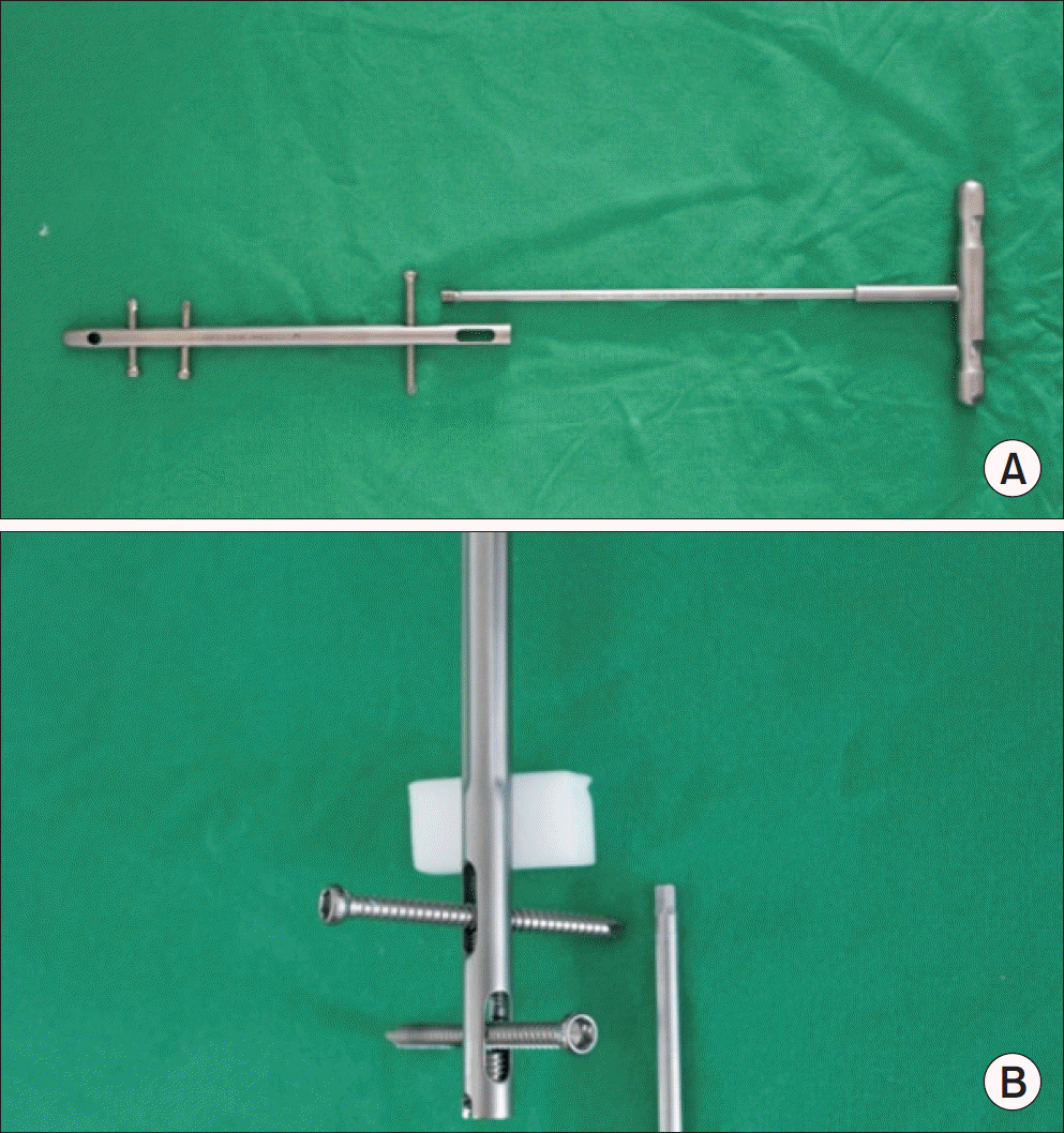
Tibiotalocalcaneal arthrodesis is done with Dyna Locking Ankle Nail (U&I Corporation). (A) Anteropostieror view. (B) Oblique view.

Figure 2.
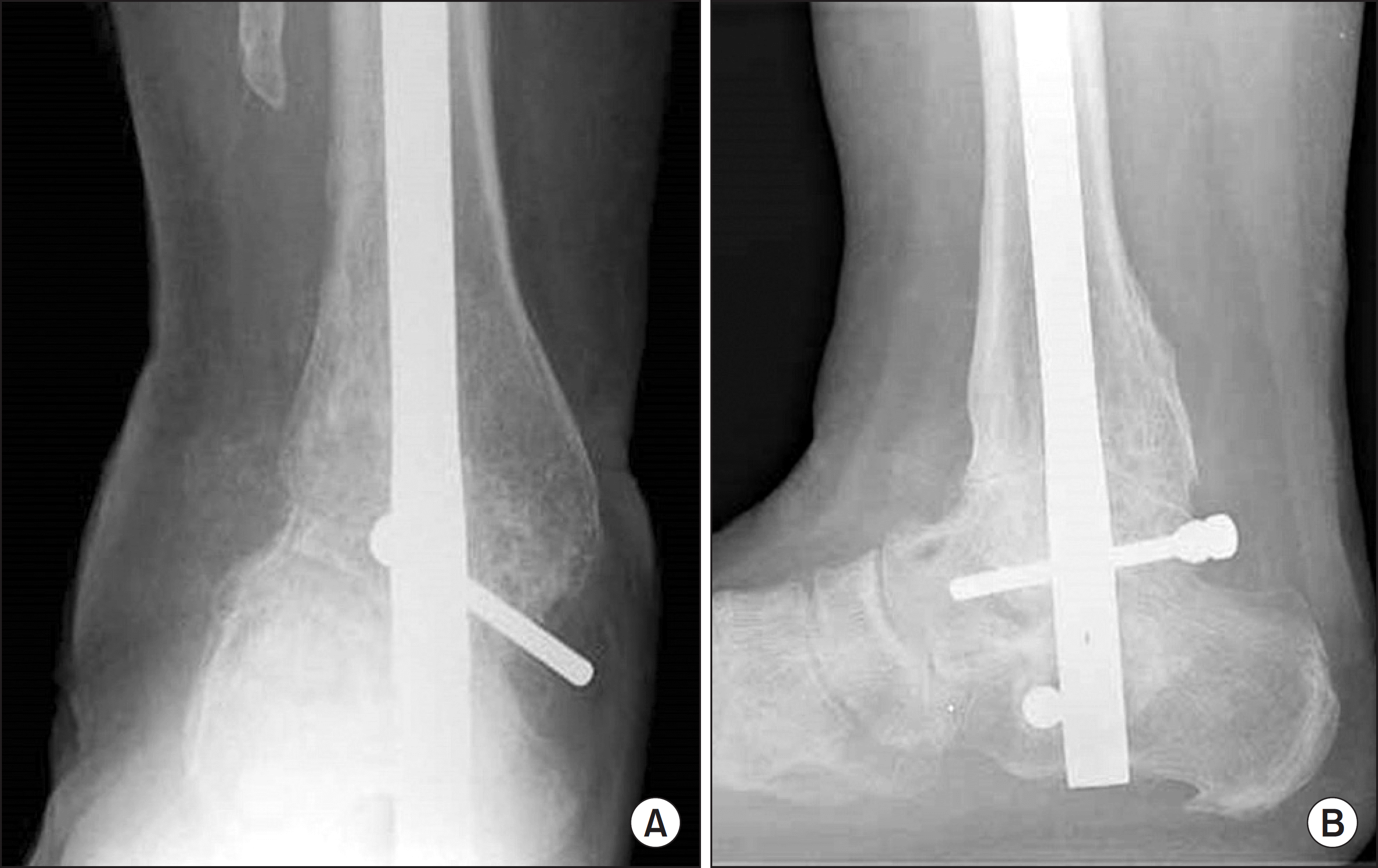
Radiographs show successful fusion trabeculae crossing the fusion site on plain anteroposterior (A) and lateral (B) radiographs.

Figure 3.
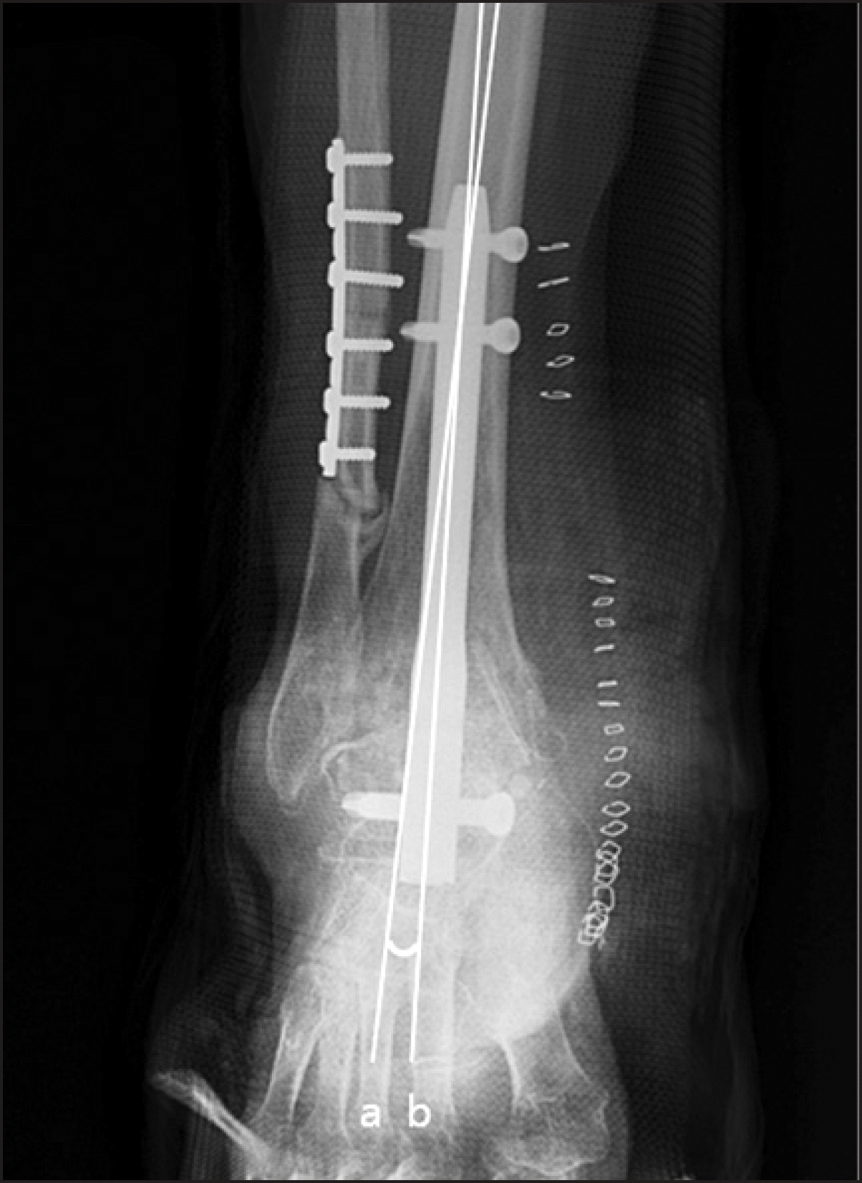
Nail-tibial angle is measured between the line bisecting the intramedullary canal of the tibia (line a) and the line bisecting (connecting) the nail (line b).

Figure 4.
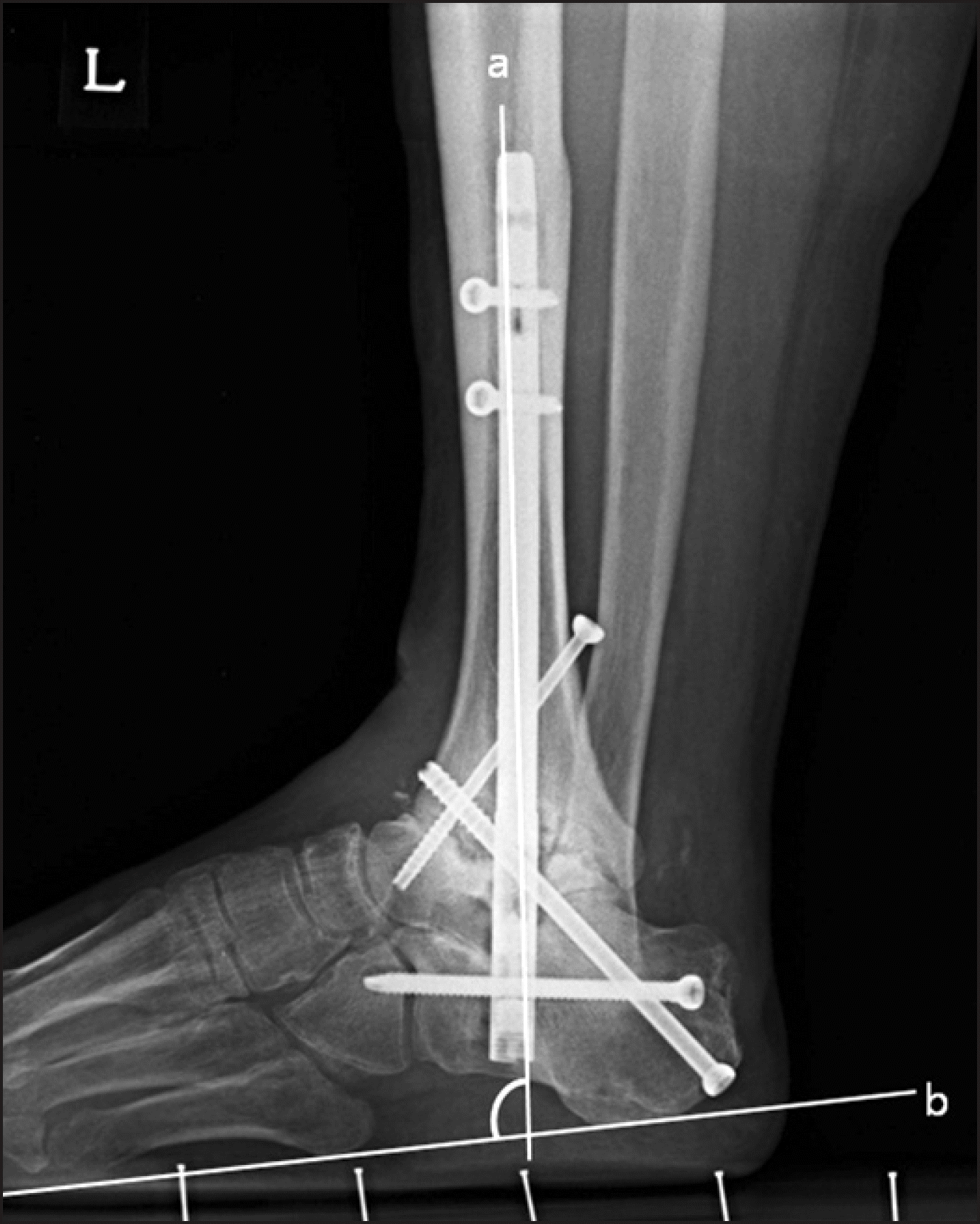
Fifth metatarsal-tibial angle is measured between the line bisecting the intramedullary canal of the tibia (line a) and the line bisecting (connecting) inferior margin of 5th metatarsus (line b).

Table 1.
Change of the Mean AOFAS and VAS Score
| Preoperative | Postoperative 6 mo | |
|---|---|---|
| AOFAS score | ||
| Pain | 12 | 32 |
| Function | 24 | 41 |
| Alignment | 5 | 8 |
| VAS score | 8.2 | 3.8 |
Table 2.
Logistic Regression Analysis between Groups of Postoperative 6 Months AOFAS Score over 75 and below 75


 PDF
PDF ePub
ePub Citation
 Print
Print


 XML Download