

 PDF
PDF ePub
ePub Citation
 Print
Print


Abstract
Purpose
We compared plain radiographs with computed tomography (CT) images to evaluate the usefulness of preoperative CT in acute ankle malleolar fracture in terms of accuracy of diagnosis and planning of operative strategy.
Materials and Methods
A retrospective analysis was conducted on 210 cases of malleolar fracture treated at our institute for which plain radiograph and CT were obtained preoperatively. Observers had reviewed plain radiographs and recorded fracture classification, anatomical diagnosis, extent and configuration of fractures and then subsequently reviewed CT images. Records from each image were compared and information regarding the differences in fractures was assessed.
References
1. Irwin TA, Lien J, Kadakia AR. Posterior malleolus fracture. J Am Acad Orthop Surg. 2013; 21:32–40.

2. Phisitkul P, Ebinger T, Goetz J, Vaseenon T, Marsh JL. Forceps reduction of the syndesmosis in rotational ankle fractures: a cadaveric study. J Bone Joint Surg Am. 2012; 94:2256–61.
3. Magid D, Michelson JD, Ney DR, Fishman EK. Adult ankle fractures: comparison of plain films and interactive two- and three-dimensional CT scans. AJR Am J Roentgenol. 1990; 154:1017–23.
4. Büchler L, Tannast M, Bonel HM, Weber M. Reliability of radiologic assessment of the fracture anatomy at the posterior tibial plafond in malleolar fractures. J Orthop Trauma. 2009; 23:208–12.
5. Ferries JS, DeCoster TA, Firoozbakhsh KK, Garcia JF, Miller RA. Plain radiographic interpretation in trimalleolar ankle fractures poorly assesses posterior fragment size. J Orthop Trauma. 1994; 8:328–31.
6. Haraguchi N, Haruyama H, Toga H, Kato F. Pathoanatomy of posterior malleolar fractures of the ankle. J Bone Joint Surg Am. 2006; 88:1085–92.
7. Black EM, Antoci V, Lee JT, Weaver MJ, Johnson AH, Susarla SM, et al. Role of preoperative computed tomography scans in operative planning for malleolar ankle fractures. Foot Ankle Int. 2013; 34:697–704.
Figure 1.
(A) No definite posterior malleolus fracture was seen in plain radiograph. (B) Posterior malleolar fracture gap was revealed by computed tomography.
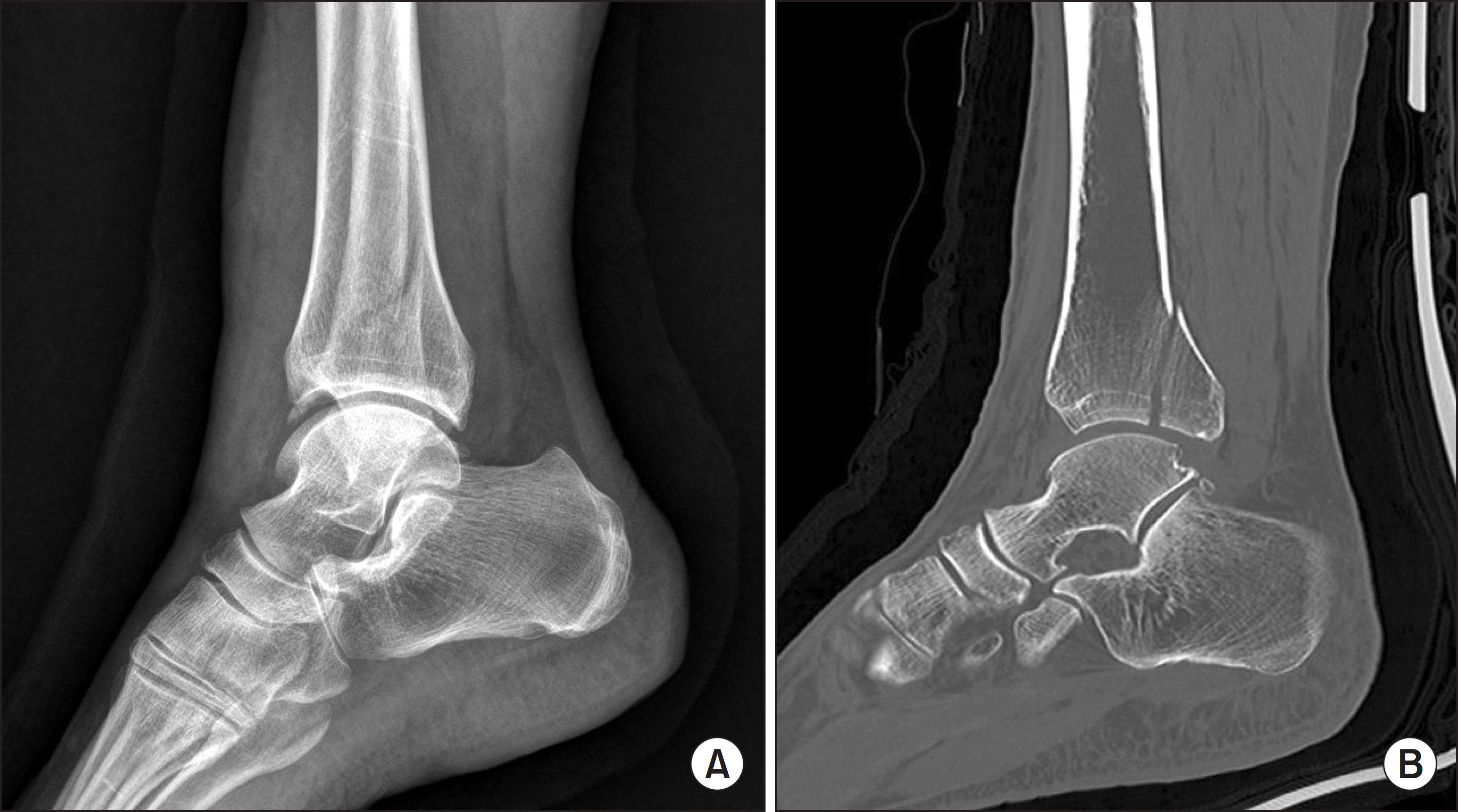
Figure 2.
(A) Other fracture line was missed by initial plain radiograph. (B) Additional Chaput tubercle fracture was found after reviewing the axial view of computed tomography image. (C) Chaput tubercle fragment was fixed with one 2.7 mm cortical screw.
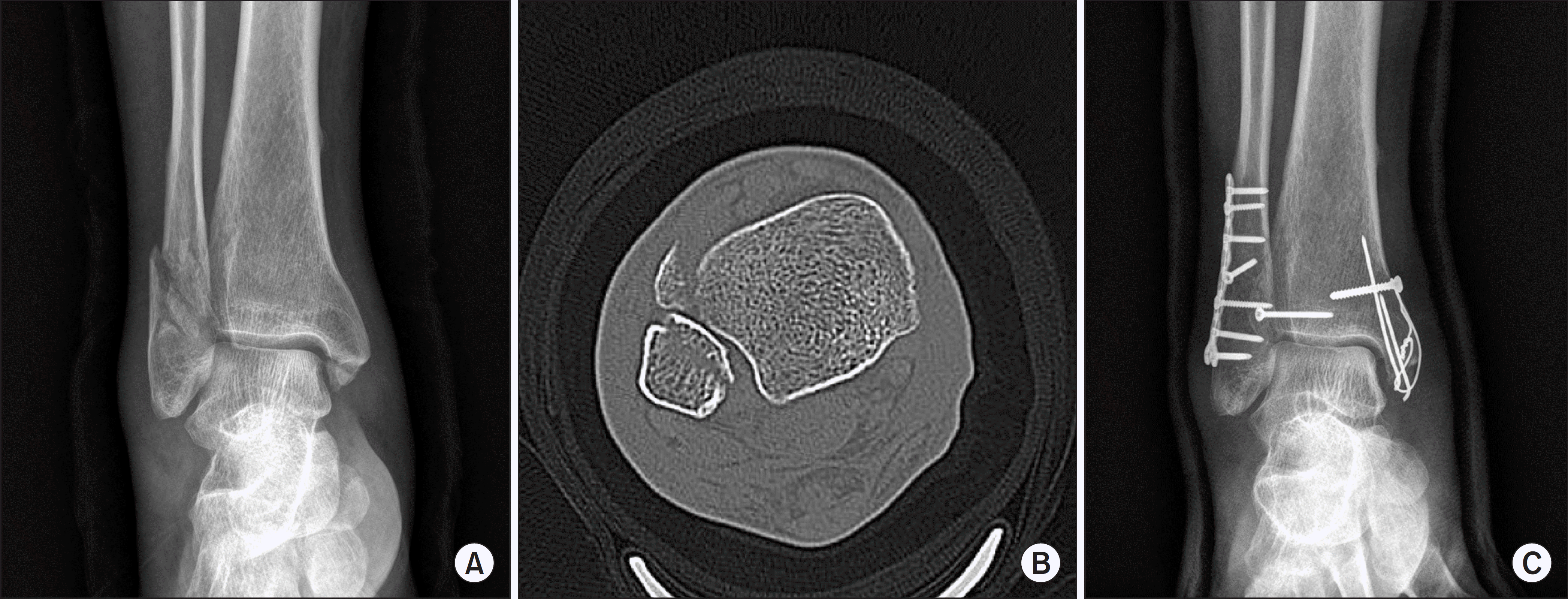
Table 1.
Changes of Lauge-Hansen Classification after Reviewing Computed Tomography (CT) Images
Table 2.
Changes of Anatomic Diagnosis of Malleolar Fractures after Reviewing Computed Tomography (CT) Images
Table 3.
Additional Findings after Reviewing Computed Tomography (CT) Images
 XML Download