

 PDF
PDF ePub
ePub Citation
 Print
Print


Abstract
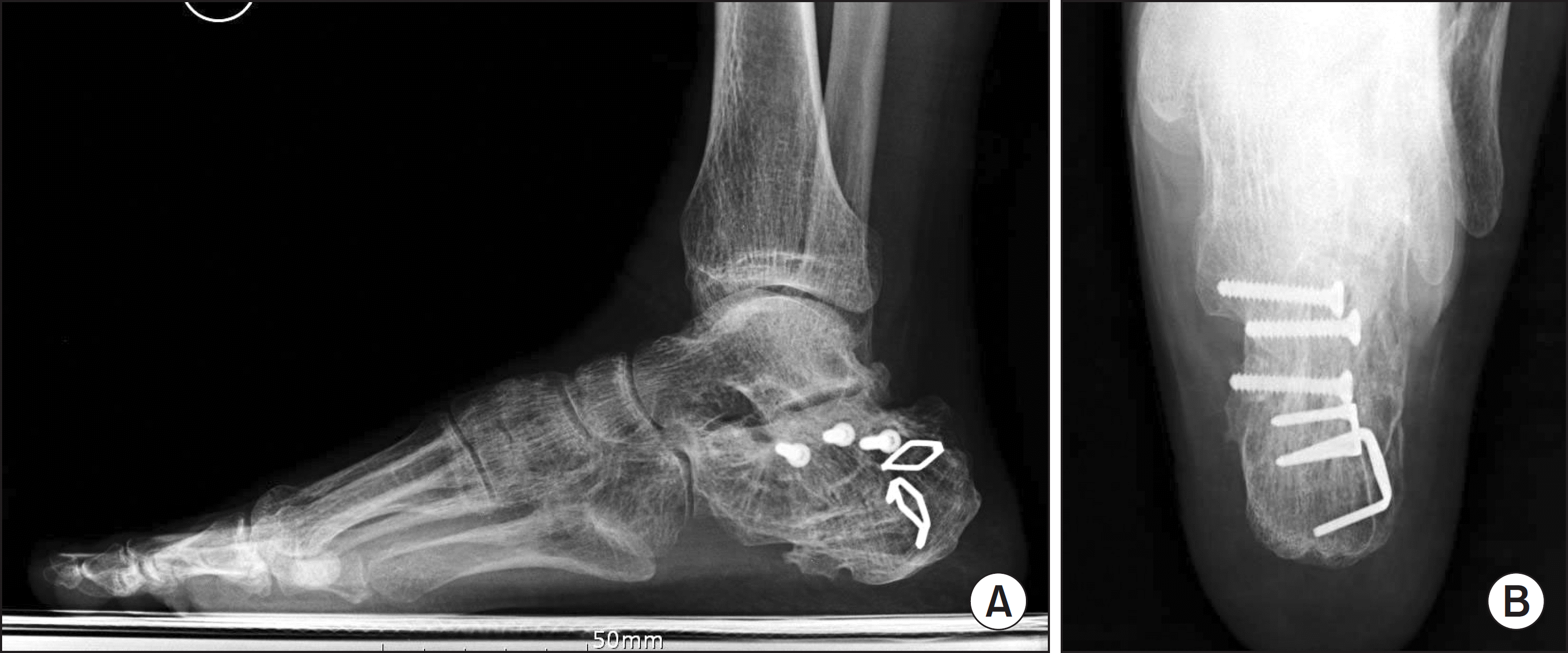
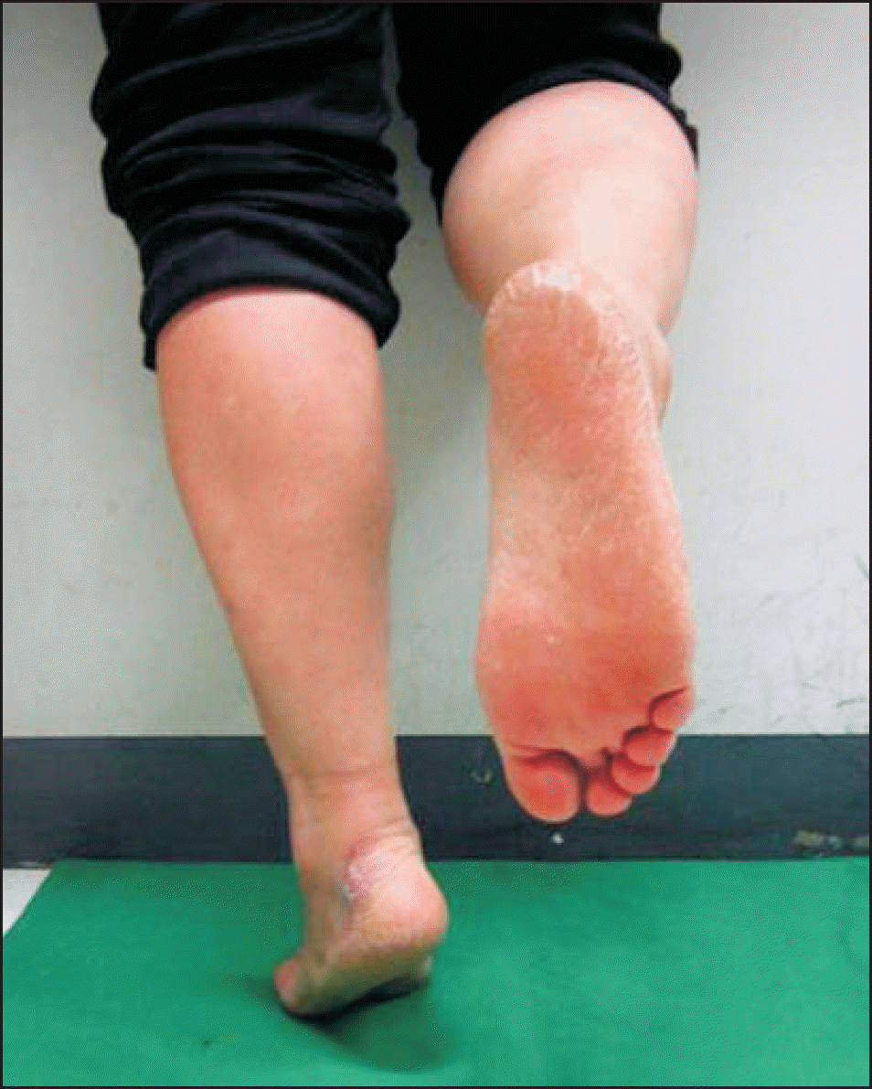
Inappropriate treatment for calcaneus fracture may result in malunion causing long-lasting pain and functional deficits. When such complications occur, the ideal principle of management is preserving congruence and motion of adjacent joints. For three patients with calcaneus fracture malunion, subtalar joint-preserving surgery using exostectomy and corrective osteotomy was performed, and satisfactory outcomes were achieved postoperatively.
References
1. Banerjee R, Saltzman C, Anderson RB, Nickisch F. Management of calcaneal malunion. J Am Acad Orthop Surg. 2011; 19:27–36.

2. Lim EV, Leung JP. Complications of intraarticular calcaneal fractures. Clin Orthop Relat Res. 2001; 391:7–16.
3. Carr JB. Mechanism and pathoanatomy of the intraarticular calcaneal fracture. Clin Orthop Relat Res. 1993; 290:36–40.
4. Yu GR, Hu SJ, Yang YF, Zhao HM, Zhang SM. Reconstruction of calcaneal fracture malunion with osteotomy and subtalar joint salvage: technique and outcomes. Foot Ankle Int. 2013; 34:726–33.
5. Aktuglu K, Aydogan U. The functional outcome of displaced intraarticular calcaneal fractures: a comparison between isolated cases and polytrauma patients. Foot Ankle Int. 2002; 23:314–8.
6. Mann RA, Baumgarten M. Subtalar fusion for isolated subtalar disorders. Preliminary report. Clin Orthop Relat Res. 1988; (226):260–5.
7. Savory KM, Wülker N, Stukenborg C, Alfke D. Biomechanics of the hindfoot joints in response to degenerative hindfoot arthrodeses. Clin Biomech (Bristol, Avon). 1998; 13:62–70.
8. Rammelt S, Grass R, Zwipp H. Joint-preserving osteotomy for malunited intraarticular calcaneal fractures. J Orthop Trauma. 2013; 27:e234–8.
9. Chen YJ, Huang TJ, Hsu KY, Hsu RW, Chen CW. Subtalar dis-tractional realignment arthrodesis with wedge bone grafting and lateral decompression for calcaneal malunion. J Trauma. 1998; 45:729–37.
10. Myerson M, Quill GE Jr. Late complications of fractures of the calcaneus. J Bone Joint Surg Am. 1993; 75:331–41.
Figure 1.
Preoperative standing lateral radiograph of affected side (A) shows decreased talocalcaneal height, talar declination angle, talocalcaneal angle, and calcaneal pitch. Sagittal (B) and coronal (C) images of preoperative computed tomography show compressed posterior subtalar facet, widened heel with lateral exostosis, and incongruity of subtalar joint.
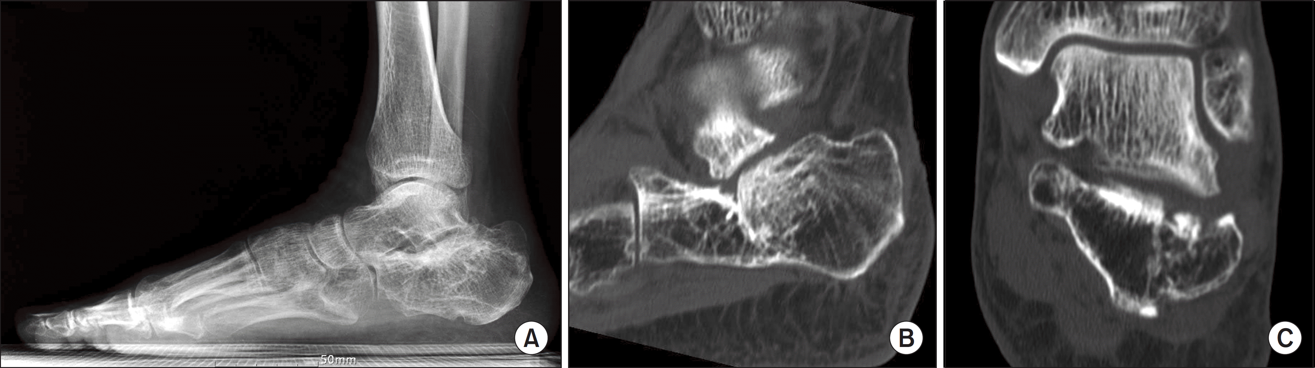
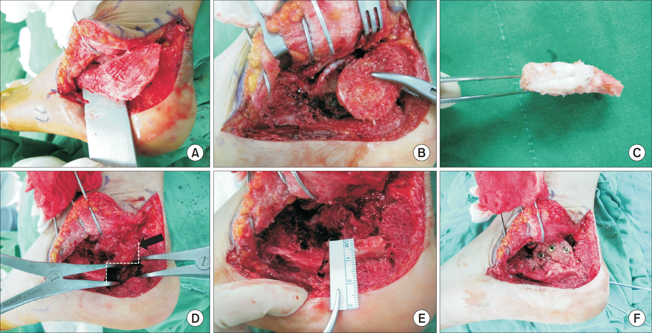
Figure 2.
Lateral wall exostectomy and corrective osteotomy were performed as subtalar joint-preserving surgery. (A) Lateral wall exostectomy was performed with an osteotome and the excised bone fragment was saved. (B) We inspected the subtalar joint and separated the depressed posterior articular facet fragment along the primary fracture line. (C) And debridement of the excised posterior facet was done. (D) With laminar spreader, calcaneus body was distracted to reestablish the height following stepped corrective osteotomy (arrow). (E) Bone graft was done with excised lateral wall fragment. (F) Then, malunited posterior facet fragment was reduced and fixed internally.
 XML Download