Journal List > J Korean Foot Ankle Soc > v.19(1) > 1043345

Lee, Kang, Hwang, Noh, and Lee: Usefulness of Treatment with 6.5 mm Cancellous Screw and Steinmann Pin Fixation for Calcaneal Joint Depression Fracture

Abstract

Purpose:

To report the radiographic and clinical results of 6.5 mm cancellous full threaded buttress screw or Steinmann pin fixation to maintain a reduction of calcaneal posterior facet depression fracture.

Materials and Methods:

From June 2009 to June 2012, 50 consecutive cases with calcaneal joint depression fracture that underwent open reduction and screw or pin fixation were enrolled in this study. A 6.5 mm cancellous full threaded screw was inserted from the posteroinferior aspect of the calcaneal tuberosity to the posterior facet (group A) or Steinman pin was inserted from the posterosuperior aspect of the calcaneal tuberosity to the calcaneocuboidal joint (group B). Both preoperative and postoperative Bohler and Gissane angles were measured radiographically, and American Orthopaedic Foot and Ankle Society (AOFAS) ankle-hindfoot scale on the final follow-up were assessed.

Results:

The mean age of patients was 44.1 years, and the mean follow-up period was 27.2 months. According to the Sanders classification, 28 cases were type II and 22 cases were type III. In Sanders type II, Bohler and Gissane angles improved significantly from 10.1° and 126.2° preoperatively to 27.2° and 117.1°, respectively, in the immediate postoperative radiograph, and at the final follow-up, 26.6° and 118.6°, respectively. In Sanders type III, Bohler and Gissane angles improved significantly from 5.0° and 129.8° to 29.9° and 119.3°, respectively, in the immediate postoperative radiograph, and 26.9° and 120.2° at the final follow-up. All cases achieved bony union, and the average period until complete union was 13.3 weeks. AOFAS ankle-hindfoot scale was 82.6 in Sanders type II and 77.3 in Sanders type III at the final follow-up.

Conclusion:

A 6.5 mm cancellous full threaded buttress screw or Steinman pin fixation is a noninvasive treatment method with a merit of being able to maintain the bearing capacity of the posterior facet comparable to plate fixation.

REFERENCES

1.Cave EF. Fracture of the os calcis--the problem in general. Clin Orthop Relat Res. 1963. 30:64–6.
2.Fitzgibbons TC., McMullen ST., Mormino MA. Fractures and dislocations of the calcaneus. Bucholz RW, editor. editor.Rockwood and Green’s factures in adults. 5th ed.Philadelphia: Lippincott Williams & Wilkins;2001. p. 2133–79.
3.Murphy GA. Fractures and dislocations of foot. Canale ST, Campbell WC, editors. editors.Campbell’s operative orthopaedics. 10th ed.St. Louis: Mosby;2003. p. 4231–83.
4.Tscherne H., Schatzker J. Major fractures of the pilon, the talus and the calcaneus: current concepts of treatment. Berlin: Springer-Verlag;1993. p. 153–74.
5.Abidi NA., Dhawan S., Gruen GS., Vogt MT., Conti SF. Wound-healing risk factors after open reduction and internal fixation of calcaneal fractures. Foot Ankle Int. 1998. 19:856–61.
crossref
6.Benirschke SK., Sangeorzan BJ. Extensive intraarticular fractures of the foot. Surgical management of calcaneal fractures. Clin Orthop Relat Res. 1993. 292:128–34.
7.Barei DP., Bellabarba C., Sangeorzan BJ., Benirschke SK. Fractures of the calcaneus. Orthop Clin North Am. 2002. 33:263–85.
crossref
8.Stephenson JR. Surgical treatment of displaced intraarticular fractures of the calcaneus. A combined lateral and medial ap-proach. Clin Orthop Relat Res. 1993. 290:68–75.
9.Essex-Lopresti P. The mechanism, reduction technique, and results in fractures of the os calcis. Br J Surg. 1952. 39:395–419.
crossref
10.Letournel E. Open reduction and internal fixation of calcaneus fractures. Spiegel PG, editor. editor.Topics in orthopedic trauma. 1st ed.Baltimore: University Parkpress;1984. p. 173–92.
11.Letournel E. Open treatment of acute calcaneal fractures. Clin Orthop Relat Res. 1993. 290:60–7.
crossref
12.Zwipp H., Tscherne H., Thermann H., Weber T. Osteosynthesis of displaced intraarticular fractures of the calcaneus. Results in 123 cases. Clin Orthop Relat Res. 1993. 290:76–86.
13.Sanders R., Fortin P., DiPasquale T., Walling A. Operative treatment in 120 displaced intraarticular calcaneal fractures. Results using a prognostic computed tomography scan classification. Clin Orthop Relat Res. 1993. 290:87–95.
14.Ross SD., Sowerby MR. The operative treatment of fractures of the os calcis. Clin Orthop Relat Res. 1985. 199:132–43.
crossref
15.Thordarson DB., Krieger LE. Operative vs. nonoperative treatment of intra-articular fractures of the calcaneus: a prospective randomized trial. Foot Ankle Int. 1996. 17:2–9.
crossref
16.Basile A. Operative versus nonoperative treatment of displaced intra-articular calcaneal fractures in elderly patients. J Foot Ankle Surg. 2010. 49:25–32.
crossref
17.Robb CA., Deans V., Iqbal MJ., Cooper JP. Comparison of non-operative and surgical treatment of displaced calcaneal fractures. The Foot. 2007. 17:169–73.
crossref
18.Palmer I. The mechanism and treatment of fractures of the calcaneus; open reduction with the use of cancellous grafts. J Bone Joint Surg Am. 1948. 30:2–8.
19.Maxfield JE., McDermott FJ. Experiences with the Palmer open reduction of fractures of the calcaneus. J Bone Joint Surg Am. 1955. 37:99–106.
crossref
20.McReynold IS. The case on operative treatment of the os calcis. Leach RE, Hoaglund FT, Riseborough EJ, editors. editors.Controversies in orthopedic surgery. Philadelphia: Sanders;1982. p. 232–54.
21.Levine DS., Helfet DL. An introduction to the minimally invasive osteosynthesis of intra-articular calcaneal fractures. Injury. 2001. 32(Suppl 1):SA51-4.
crossref
22.Schepers T., Schipper IB., Vogels LM., Ginai AZ., Mulder PG., Heetveld MJ, et al. Percutaneous treatment of displaced intra-articular calcaneal fractures. J Orthop Sci. 2007. 12:22–7.
crossref
23.Widen A. Fractures of the calcaneus: a clinical study with special reference to the technique and results of open reduction. Acta Chir Scand Suppl. 1954. 188:1–119.
24.Lee MJ., Sohn SK., Lee KY., Kim SS., Kang MS., Kim HJ, et al. Open reduction and internal fixation with AO calcaneal plate for displaced intra-articular calcaneal fracture. J Korean Fract Soc. 2010. 23:303–9.
crossref
25.Moon JS., Lee WC. A comparison of extensile lateral approach and sinus tarsi approach for the sanders type II calcaneal fracture. J Korean Fract Soc. 2009. 22:13–8.
crossref
26.Lee YT., Oh HC., Yoon HK., Jang JW., Jang KJ. Comparison of F calcaneal plate and locking calcaneal plate fixation using an lateral extensile approach to intra-articular calcaneal fractures. J Korean Foot Ankle Soc. 2012. 16:175–80.

Figure 1.
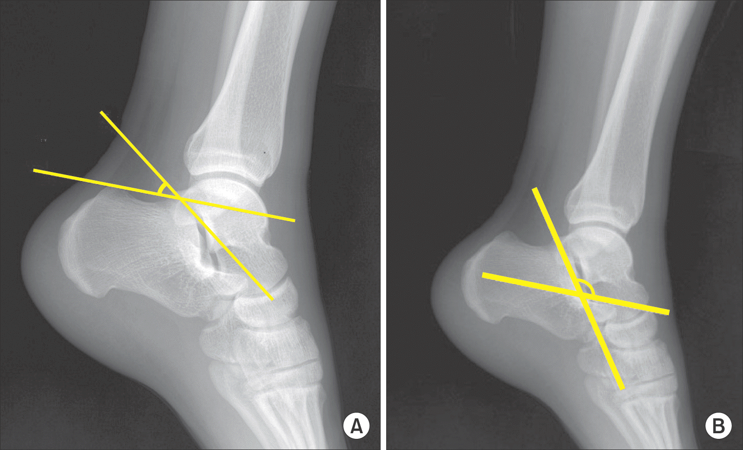
Measurement of Bohler and Gissane angles. (A) Bohler angle is measured by the intersection of a line drawn from most cephalic point on tuberosity to highest point of posterior facet with a line from highest point of posterior facet to most cephalic part of posterior process of calcaneus. (B) Gissane angle is measured by the intersection of a line drawn from the highest point of the posterior articular facet to the highest point of the posterior tuberosity and a line from the former to the highest point on the anterior articular facet.
jkfas-19-11f1.tif
Figure 2.
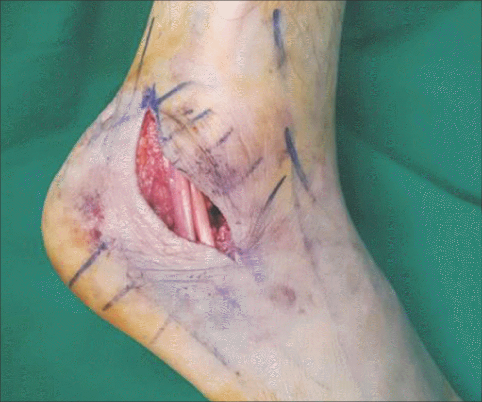
Beginning from 2.5 cm superior to lateral malleolus tip, which is the midpoint between malleolus and Achilles tendon, the incision continues obliquely and distally toward 1 cm below the lateral malleolus tip, sinus tarsi, and dorsolateral aspect of the talonavicular joint.
jkfas-19-11f2.tif
Figure 3.
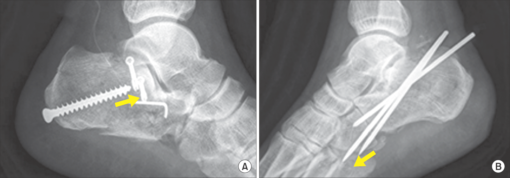
Operative method. (A) A 6.5 mm cancellous buttress screw should be inserted from posteroinferior aspect of calcaneal tuberosity to the posterior facet (arrow). (B) Steinmann pins should be inserted from posterosuperior aspect of calcaneal tuberosity to the calcaneocuboid joint (arrow).
jkfas-19-11f3.tif
Table 1.
Demographic Data of Patients
Variable Group A Group B p-value
Number of patients 32 18
Mean age (yr) 44.5 (21∼63) 43.5 (24∼61) 0.593
Gender (male) 22 10 0.869
Affected foot (right) 14 9 0.612
Smoker 10 4 0.245

Values are presented as number or mean (range). Group A: a 6.5 mm cancellous full threaded screw inserted from the posteroinferior aspect of the calcaneal tuberosity to the posterior facet Group B: Steinman pin inserted from the posterosuperior aspect of the calcaneal tuberosity to the calcaneocuboidal joint.

Table 2.
Comparison between Preoperative and Postoperative Bohler and Gissane Angles in Sanders Type II Calcaneus Fractures (n=28)
Angle Preoperative Postoperative Final follow-up p-value* p-value
Bohler angle 10.1°±4.4° 27.2°±2.2° 26.6°±2.5° 0.009 0.845
Gissane angle 126.2°±5.5° 117.1°±3.2° 118.6°±4.4° 0.011 0.385

* Values are presented as mean±standard deviation. Comparison between preoperative and postoperative Bohler and Gissane angles.

Comparison between postoperative and final follow-up Bohler and Gissane angles.

Table 3.
Comparison between Preoperative and Postoperative Bohler and Gissane Angles in Sanders Type III Calcaneus Fractures (n=22)
Angle Preoperative Postoperative Final follow-up p-value* p-value
Bohler angle 5.0°±3.4° 29.9°±3.1° 26.9°±3.0° 0.001 0.557
Gissane angle 129.8°±9.5° 119.3°±8.7° 120.2°±12.0° 0.004 0.985

Values are presented as mean±standard deviation. *Comparison between preoperative and postoperative Bohler and Gissane angles.

Comparison between postoperative and final follow-up Bohler and Gissane angles.

Table 4.
Comparison between Postoperative and Final Follow-up Bohler Angles for Group A and B in Sanders Classification Type II and III Calcaneus Fractures
Type Group A (n=32) Group B (n=18)
Postoperative Final follow-up Postoperative Final follow-up p-value
Type II (n=28) 28.7°±2.5° 27.8°±2.2° 24.5°±2.1° 24.4°±1.8° 0.727
Type III (n=22) 32.9°±4.7° 28.7°±3.5° 24.7°±3.9° 23.7°±7.5° 0.825

Values are presented as mean±standard deviation. Group A: a 6.5 mm cancellous full threaded screw inserted from the posteroinferior aspect of the calcaneal tuberosity to the posterior facet; Group B Steinman pin inserted from the posterosuperior aspect of the calcaneal tuberosity to the calcaneocuboidal joint.

Table 5.
Comparison between Postoperative and Final Follow-up Gissane Angle of Group A and B in Sanders Classification Type II and III Calcaneus Fractures
Type Group A (n=32) Group B (n=18)
Postoperative Final follow-up Postoperative Final follow-up p-value
Type II (n=28) 117.2°±1.9° 118.9°±2.1° 116.8°±2.0° 118.1°±1.8° 0.862
Type III (n=22) 118.9°±2.0° 120.7°±2.5° 120.0°±6.0° 119.3°±5.8° 0.740

Values are presented as mean±standard deviation. Group A: a 6.5 mm cancellous full threaded screw inserted from the posteroinferior aspect of the calcaneal tuberosity to the posterior facet; Group B Steinman pin inserted from the posterosuperior aspect of the calcaneal tuberosity to the calcaneocuboidal joint.

Table 6.
Comparison of AOFAS Ankle-Hindfoot Scale between Group A and B in Sanders Classification Type II and III Calcaneus Fractures
Type Group A (n=32) Group B (n=18) p-value
Type II (n=28) 82.5±4.6 82.7±5.8 0.960
Type III (n=22) 76.2±3.7 79.3±4.2 0.796

Values are presented as mean±standard deviation. Group A: a 6.5 mm cancellous full threaded screw inserted from the posteroinferior aspect of the calcaneal tuberosity to the posterior facet; Group B: Steinman pin inserted from the posterosuperior aspect of the calcaneal tuberosity to the calcaneocuboidal joint. AOFAS: American Orthopaedic Foot and Ankle Society.

TOOLS
Similar articles